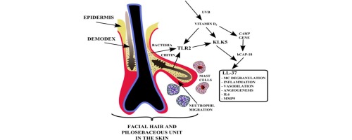
Phân tích hệ vi sinh vật của Demodex spp. thu được từ da của những người mắc ETR, PPR và da bình thường cho thấy sự đa dạng vi sinh vật khác nhau tùy thuộc vào tình trạng da của vật chủ. Người ta cho rằng hệ vi sinh vật liên quan đến Demodex spp. có thể bị ảnh hưởng bởi thành phần bã nhờn khác nhau trong da bị bệnh trứng cá đỏ. Nghiên cứu gần đây chỉ ra vai trò của vi khuẩn liên quan đến Demodex spp. là Bacillus oleronius trong việc kích thích các phản ứng viêm trong PPR, ETR và OR. B. oleronius được phân lập từ D. folliculorum lấy từ mặt của một bệnh nhân bị bệnh trứng cá đỏ dạng sẩn mụn mủ và vi khuẩn này nhạy cảm với tetracycline, doxycycline và minocycline, những loại thuốc thường được sử dụng thành công trong điều trị bệnh trứng cá đỏ. Đặc biệt đáng quan tâm là các protein 62‐ và 83 kDa có nguồn gốc từ B. oleronius vì chúng dường như kích hoạt phản ứng miễn dịch đáng kể trong huyết thanh của những người mắc bệnh trứng cá đỏ khi so sánh với nhóm đối chứng. Tiếp xúc với protein B. oleronius gây ra tỷ lệ cao của quá trình hóa hướng bạch cầu trung tính và phản ứng viêm do BCĐNTT trung gian liên quan đến IL‐8, MMP9, TNF‐α và cathelicidin.
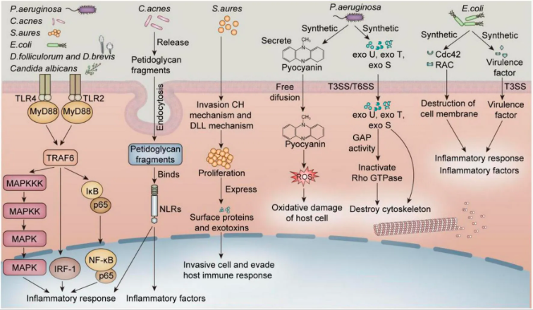
Hình 13. Vi sinh vật phổ biến gây viêm bờ mi thông qua nhiều cơ chế tiềm năng khác nhau: Nội bào ‐ Nội bào là quá trình tế bào hấp thụ các chất bên ngoài (như chất dinh dưỡng, phân tử, mầm bệnh), được bao bọc bởi màng tế bào để tạo thành các túi nội bào & vận chuyển đến vị trí khác nhau bên trong tế bào. Staphylococcus aureus và C. acnes gây ra phản ứng viêm bằng cách kích hoạt con đường TLR2(4)/MYD88/NF‐KB trong tế bào sừng và tế bào tuyến bã. S. aureus xâm nhập vào tế bào và né tránh phản ứng miễn dịch của vật chủ & đã được chứng minh là có mặt trong nguyên bào sợi, tế bào sừng và tế bào nội mô.
Phản ứng miễn dịch do P. aeruginosa và E. coli gây ra cũng có thể làm tổn thương các tế bào nội mô và tế bào sừng. Các nghiên cứu cũng chỉ ra Demodex spp. gây viêm bằng cách tăng biểu hiện TLR2 trong tế bào sừng. Các tế bào ở rìa mi mắt gồm tế bào sừng, biểu mô, tuyến bã nhờn xung quanh nang lông mi và tế bào tuyến mồ hôi, cùng nhiều loại tế bào khác. Do đó, các cơ chế này có thể liên quan chặt chẽ đến viêm bờ mi.
Nguồn: https://www.frontiersin.org/journals/medicine/articles/fmed.2026.1801375/full
Vai trò tiềm tàng của protein B. oleronius trong sự phát triển của loét giác mạc gần đây đã được nghiên cứu bằng cách cho các tế bào biểu mô giác mạc (hTCEpi) tiếp xúc với protein Bacillus và điều này dẫn đến sự gia tăng của một số cytokine gây viêm bao gồm IL‐6, IL‐8, IL‐1β và TNF‐α. Trong một nghiên cứu riêng biệt sử dụng cùng dòng tế bào, sự gia tăng đáng kể của MMP3 và MMP9 đã được quan sát thấy ở các tế bào tiếp xúc với B. oleronius , do đó dẫn đến một lời giải thích khả dĩ cho sự hình thành loét giác mạc và sẹo giác mạc ở những người mắc bệnh trứng cá đỏ ở mắt.
Hiện nay có một số phương pháp điều trị bệnh rosacea, tuy nhiên chúng thường chỉ điều trị các triệu chứng riêng biệt chứ không phải nguyên nhân cơ bản. Các loại thuốc bôi ngoài da như axit azelaic và metronidazole đã cho thấy hiệu quả trong điều trị PPR nhờ đặc tính chống viêm và kháng khuẩn của chúng. Thuốc tetracycline đường uống đã cho thấy thành công trong điều trị bệnh trứng cá đỏ mặc dù chưa rõ liệu điều này là do đặc tính kháng khuẩn của nó hay do khả năng ức chế hoạt động của protease KLK5 và MMP, do đó làm giảm viêm. Vì thuốc kháng sinh tetracycline không có tác dụng chống lại Demodex spp., nên có thể thuốc này đang tác động lên một yếu tố vi khuẩn, do đó làm giảm viêm trên da bị trứng cá đỏ. Mặt khác, điều trị bằng đường uống với liều doxycycline dưới mức kháng khuẩn, dùng một lần mỗi ngày dưới dạng viên nang 40 mg (30 mg giải phóng tức thì và 10 mg hạt giải phóng chậm) đã được chứng minh là có hiệu quả tương đương với liều doxycycline 100 mg (kháng khuẩn) trong việc giảm viêm ở những người mắc bệnh trứng cá đỏ và liệu trình điều trị này hiện đã được FDA Mỹ phê duyệt để điều trị bệnh trứng cá đỏ.
Dầu cây trà (TTO) là một chất diệt Demodex spp. đã được biết đến và đã chứng minh tác dụng tích cực trong việc giảm nhiễm Demodex và viêm ở bệnh nhân bị viêm bờ mi. Ivermectin là một loại thuốc chống KST phổ rộng cũng có một số đặc tính chống viêm.
Gần đây, việc sử dụng ivermectin tại chỗ (1%) đã được chứng minh là có hiệu quả trong việc giảm triệu chứng của PPR. Ivermectin đường uống đã có kết quả lâm sàng thành công trong điều trị nhiễm Demodex spp. ở bệnh nhân bị viêm bờ mi, dẫn đến giảm mật độ Demodex spp. và cải thiện triệu chứng lâm sàng. Trong nghiên cứu này và các nghiên cứu khác, việc sử dụng ivermectin toàn thân không loại bỏ hoàn toàn Demodex.
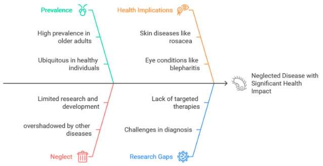
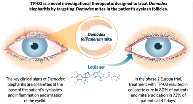
Hình 14. Hiểu rõ tác động và thách thức của bệnh da do Demodex
(Nguồn: https://link.springer.com/article/10.1007/s15010‐025‐02521‐z/figures/5)
Tuy nhiên, mối tương quan giữa việc giảm các triệu chứng lâm sàng được quan sát và giảm nhiễm Demodex spp. ở bệnh nhân được điều trị bằng ivermectin củng cố lập luận rằng Demodex đóng vai trò quan trọng trong các biểu hiện của OR và các triệu chứng liên quan. Các tác giả đề xuất sử dụng ivermectin kết hợp với các liệu pháp khác để chống lại sự xâm nhiễm Demodex spp. có thể liên quan đến các triệu chứng viêm bờ mi. Thật vậy, liệu pháp kết hợp bao gồm ivermectin và metronidazole đã cho thấy kết quả lâm sàng tuyệt vời trong điều trị những người bị bệnh trứng cá đỏ trên mặt và viêm bờ mi. So sánh những người được điều trị bằng sự kết hợp của metronidazole và ivermectin so với chỉ dùng ivermectin đơn độc, cho thấy sự giảm số lượng Demodex spp. nhiều hơn ở những người được điều trị bằng liệu pháp kết hợp so với chỉ dùng ivermectin đơn độc. Đặc tính chống viêm của metronidazole được cho là làm giảm các triệu chứng viêm có thể do các kháng nguyên liên quan đến Demodex spp. gây ra và người ta cho rằng metronidazole cũng có tác dụng diệt ký sinh trùng, có thể bổ sung cho hoạt động chống Demodex của ivermectin.
Viêm da mặt không đặc hiệu
Bệnh nhân có các triệu chứng ở mặt không đặc hiệu như ngứa mặt có hoặc không kèm ban đỏ, phát ban giống viêm da tiết bã, tổn thương giống viêm da quanh miệng và tổn thương dạng sẩn mụn mủ và/ hoặc dạng mụn trứng cá mà không có giãn mao mạch, đỏ bừng hoặc mụn đầu đen được phát hiện có mật độ Demodex spp. trung bình cao hơn đáng kể.
Viêm da Demodex spp. thực tế có thể khác với bệnh trứng cá đỏ và viêm da tiết bã, như một nhóm đã báo cáo và sự hiện diện của chứng đỏ mặt, khô, bong vảy và thô ráp có hoặc không có sẩn/mụn mủ có thể là kết quả của sự phát triển quá mức của D. folliculorum. Bệnh ghẻ do Demodex ở người là do biểu hiện lâm sàng của sự nhiễm Demodex spp. ở người, vốn không có triệu chứng rõ rệt, bởi hai loài Demodex, đó là D. folliculorum và D. brevis. Cần lưu ý vai trò gây bệnh của loài đa năng này vì bệnh do Demodex ở người có thể biểu hiện dưới nhiều dạng bệnh lý khác nhau, bắt chước nhiều bệnh da liễu khác. Điều này có thể giúp chẩn đoán sớm và điều trị đúng cách, từ đó tiết kiệm thời gian và chi phí.
Kết luận
Demodex spp. có thể được tìm thấy ở người khỏe mạnh mà không gây ra bất kỳ triệu chứng lâm sàng nào. Trong những điều kiện thích hợp, khi mật độ Demodex spp. tăng lên, chúng có thể gây ra bệnh Demodicosis. Tuy nhiên, vai trò của chúng như là tác nhân gây bệnh chính trong các tình trạng bệnh lý ở người vẫn đang được tranh luận. Demodex spp., giống như các vi sinh vật ở da khác, có thể chuyển thành tác nhân gây bệnh cơ hội. Chúng có khả năng thay đổi trạng thái từ cộng sinh nếu hệ miễn dịch của vật chủ và môi trường da tạo điều kiện thuận lợi cho sự sinh sôi nảy nở của chúng. Các triệu chứng lâm sàng và mức độ nghiêm trọng của bệnh Demodicosis cũng phụ thuộc vào những điều kiện này.
Ngoài các triệu chứng “nổi tiếng” của bệnh demodicosis như vảy phấn hồngs do D. folliculorum, dạng trứng cá đỏ, dạng viêm nang lông, viêm da quanh miệng, còn có nhiều triệu chứng lâm sàng khác đã được báo cáo. Bệnh demodicosis có thể biểu hiện với các triệu chứng không đặc hiệu ở mặt như ngứa không rõ nguyên nhân, da khô từng mảng, da quá mẫn cảm, các tổn thương dạng sẩn mụn mủ và nốt sần không đặc hiệu.
Tất cả các triệu chứng này đều được phát hiện có liên quan mật thiết đến sự tăng sinh bất thường của Demodex spp. Bệnh Demodicosis là một căn bệnh khó điều trị, có thể biểu hiện với nhiều triệu chứng khác nhau, bắt chước nhiều bệnh da liễu khác. Sẽ rất có lợi cho bệnh nhân nếu các bác sĩ da liễu nhận thức rõ hơn về căn bệnh này. Ngoài ra, còn nhiều điều chưa biết về bệnh Demodicosis cần được nghiên cứu thêm.
Tài liệu tham khảo
1. House dust mite. Wikipedia Accessed 11/25/14.
2. Jarmuda S, O’Reilly N, Zaba R, et al. Potential role of Demodex mites and bacteria in the induction of rosacea. J Med Microbiol 2012;61:1504‐1510.
3. Forton FMN. Papulopustular rosacea, skin immunity and Demodex: pityriasis folliculorum as a missing link. J Eur Acad Dermatol Venereol 2012;26:19‐28.
4. NIH Human Microbiome Project defines normal bacterial makeup of the body. National Institutes of Health June 13, 2012.
5. Thoemmes MS, Fergus DJ, Urban J, Trautwein M, Dunn RR (2014) Ubiquity and Diversity of Human‐Associated Demodex Mites. PLoS ONE 9(8): e106265.
6. Marantz Henig R. Fat factors. New York Times Aug. 123, 2006.
7. Ackerman J. How bacterial affect our bodies protect our health. Scientific American May 15, 2012, pp.
8. Rusiecka‐Ziółkowska J, Nokiel M, Fleischer M. Demodex ‐ an old pathogen or a new one? Adv Clin Exp Med 2014;23:295‐298.
9. Thoemmes MS, Fergus DJ, Urban J, Trautwein M, Dunn RR (2014) Ubiquity and diversity of human‐associated Demodex mites. Public Library of Science One 9(8):e106265.
10. Jarmuda S, O’Reilly N, Zaba R, et al. Potential role of Demodex mites and bacteria in the induction of rosacea. J Medical Microbiol 2012;61:1504‐1510.
11. Zhao YE, Wu LP, Peng Y, et al. Retrospective analysis of association between Demodex infestation and rosacea. Arch Dermatol 2010;146(8):896‐902.
12. Erbagci Z, Ozgoztaosi O. The significance of Demodex folliculorum density in rosacea. Int J Dermatol 1998;37:421‐425.
13. Li J, O’Reilly N, Sheha H, et al. Correlation between ocular Demodex infestation and serum immunoreactivity to Bacillus proteins in patients with facial rosacea. Ophthalmology 2010;117:870‐877.
14. Demodex surfaces again at summer AAD meeting. National Rosacea Society Weblog Accessed 11/25/14.
15. Aylesworth R, Vance JC. Demodex folliculorum and Demodex brevis in cutaneous biopsies. J Am Acad Dermatol 1982;7:583‐9.
16. Acosta FJ, Planas L, Penneys N. Demodex mites contain immunoreactive lipase. Arch Dermatol. 1989;125:1436‐7.
17. Facey N, Delaney S, Kavanagh K, Powell FC. Mite‐related bacterial antigens stimulate inflammatory cells in rosacea. Br J Dermatol 2007;157:474‐81.
18. Spickett SG. Studies on Demodex folliculorum Simon. Parasitology 1961;51:181‐92.
19. Sędzikowska A, Bartosik K, Przydatek‐Tyrajska R, Dybicz M. Shared Makeup Cosmetics as a Route of Demodex folliculorum Infections. Acta Parasitol 2021;66:631‐7.
20. Sengbusch HG, Hauswirth JM. Prevalence of hair follicle mites, Demodex folliculorum and Demodex brevis, in a selected human population in Western New York, USA. J Med Entomol 1986;23:384‐8.
21. Özer TT, Akyürek Ö, Durmaz S. Association between Demodex folliculorum and metabolic syndrome. J Cosmet Dermatol 2020;19:3145‐9.
22. Karincaoglu Y, Seyhan ME, Bayram N, et al. Incidence of Demodex folliculorum in patients with end stage chronic renal failure. Ren Fail 2005;27:495‐9.
23. Ivy SP, Mackall CL, Gore L, et al. Demodicidosis in childhood acute lymphoblastic leukemia: An opportunistic infection occurring with immunosuppression. J Pediatr 1995;127:751‐4.
24. Fujiwara S, Okubo Y, Irisawa R, Tsuboi R. Rosaceiform dermatitis associated with topical tacrolimus treatment. J Am Acad Dermatol 2010;62:1050‐2.
25. Demirdağ HG, Özcan H, Gürsoy Ş, et al. The effects of sebum configuration on Demodex spp. Density. Turk J Med Sci 2016;46:1415‐21.
26. Thoemmes MS, Fergus DJ, Urban J, Tautwein M, Dunn RR. Ubiquity and diversity of human‐associated Demodex mite. PLoS One 2014;9: e106265.
27. Desch CE. Human hair follicle mites and forensic acarology. Exp Appl Acarol 2009;49:143‐6.
28. Esenkaya Taşbent F, Dik B. A dog related Demodex spp. infestation in a student: A rare Demodex case. Mikrobiyol Bul 2018;52:214‐20.
29. Ferreira D, Sastre N, Ravera I, Altet L, et al. Identification of a third feline Demodex species through partial sequencing of the 16S rDNA and frequency of Demodex species in 74 cats using a PCR assay. Vet Dermatol 2015;26:239.
30. Tani K, Une S, Hasegawa A, et al. Infestivity of Demodex canis to hamster skin engrafted onto SCID mice. J Vet Med Sci 2005;67:445‐8.
31. Akilov OE, KY. Association between human demodicosis and HLA class I. Clin Exp Dermatol. 2003;28:70‐3.
32. Akilov OE, Mumcuoglu KY. Immune response in demodicosis. J Eur Acad Dermatol Venereol 2004;18:440‐4.
33. Gazi U, Taylan‐Ozkan A, Mumcuoglu KY. Immune mechanisms in human and canine demodicosis: A review. Parasite Immunol 2019;41:e12673
34. Kalincaoglu Y, Tepe B, Kalayci B, Seyhan M. Pseudozoster clinical presentation of Demodex infestation after prolonged topical steroid use. Clin Exp Dermatol 2008;33:740‐2.
35. Ní Raghallaigh S, Powell FC: The cutaneous microenvironment in papulopustular rosacea. Br J Dermatol 2009;161:25.
36. Ní Raghallaigh S, Bender K, Lacey N, et al. The fatty acid profile of the skin surface lipid layer in patients with papulopustular rosacea. Br J Dermatol 2012;166:279‐87.
37. Forton F, Seys B. Density of Demodex folliculorum in rosacea: a case‐control study using standardized skin‐surface biopsy. Br J Dermatol 1993;128:650‐9.
38. Jimenez‐Acosta F, Planas L, Penneys N. Demodex mites contain immunoreactive lipase. Arch Dermatol 1989;125:1436‐7.
39. Moran EM, Foley R, Powell FC. Demodex and rosacea revisited. Clin Dermatol 2017;35:195‐200.
40. Jarmuda S, O’Reilly N, Żaba R, et al. Potential role of Demodex mites and bacteria in the induction of rosacea. J Med Microbiol 2012;61:1504‐10.
41. Fischer K, Walton S. Parasitic mites of medical and veterinary importance: Is there a common research agenda? Int J Parasitol 2014;44:955‐67.
42. O’Reilly N, Menezes N, Kavanagh K. Positive correlation between serum immunoreactivity to Demodex‐associated Bacillus proteins and erythematotelangiectatic rosacea. Br J Dermatol 2012;167:1032‐6.
43. Ionescu MA, Joly F, Bombard E, et al. TLR‐2, cathelicidin‐LL37 and kallikrein‐ 5 targeting in rosacea: In tubo, ex vivo and in vivo studies. J Am Acad Dermatol 2015;72:65.
44. Koller B, Muller Wiefel AS, Rupec R, et al. Chitin modulates innate immune responses of keratinocytes. PLoS One 2011;6:e16594.
45. Yamasaki K, Kanada K, Macleod DT, et al. TLR2 expression is increased in rosacea and stimulates enhanced serine protease production by keratinocytes. J Invest Dermatol 2011;131:688‐97.
46. Casas C, Paul C, Lahfa M, et al. Quantification of Demodex folliculorum by PCR in rosacea and its relationship to skin innate immune activation. Exp Dermatol 2012;21:906‐10.
47. Erbagci Z, Ozgoztasi O. The significance of Demodex folliculorum density in rosacea. Int J Dermatol 1998;37:421‐5.
48. Karincaoglu Y, Bayram N, Aycan O, Esrefoglu M. The clinical importance of Demodex folliculorum presenting with nonspecific facial signs and symptoms. J Dermatol 2004;31:618‐26.
49. Aktaş Karabay E, Aksu Çerman A. Demodex folliculorum infestations in common facial dermatoses: Acne vulgaris, rosacea, seborrheic dermatitis. An Bras Dermatol 2020;95:187‐93.
50. Forton F, De Maertelaer V. Rosacea‐like demodicosis and papulopustular rosacea may be two phenotypes of the same disease, and pityriasis folliculorum may be their precursor: Response to the comment of Tatu. J Eur Acad Dermatol Venereol 2019;33:e47‐8.
51. Akçınar UG, Ünal E, Doğruman Al F. Demodex spp. as a possible aetiopathogenic factor of acne and relation with acne severity and type. Postepy Dermatol Alergol 2018;35:174‐81.
52. Yun CH, Yun JH, Baek JO, et al. Demodex Mite Density Determinations by Standardized Skin Surface Biopsy (SSSB) and Direct Microscopic Examination (DME) and their relations with clinical types and distribution patterns. Ann Dermatol 2017;29:137‐42.
53. Yasak Guner R, Tosun M, Akyol M, Hayta SB. Demodex infestation as a cause of sensitive skin in a dermatology outpatient clinic. J Cosmet Dermatol. 2021. May 22. Online ahead of print.
54. Douglas A, Zaenglein AL. A case series of demodicosis in children. Pediatr Dermatol 2019;36:651‐4.
55. Forton FMN, De Maertelaer V. Rosacea and Demodicosis: Little‐known diagnostic signs and symptoms. Acta Derm Venereol 2019;99:47‐52.
56. Helou W, Avitan‐Hersh E, Bergman R. Demodex folliculitis of the scalp: Clinicopathological study of an uncommon entity. Am J Dermatopathol 2016;38:658‐63.
57. Bachmeyer C, Moreno‐Sabater A. Demodex folliculitis. CMAJ 2017;189:E865.
58. Schaller M, Sander CA, Plewig G. Demodex abscesses: Clinical and therapeutic challenges. J Am Acad Dermatol 2003;49:S272‐274.
59. Lee JY, Hsu CH. Granulomatous rosacea‐ like demodicidosis. Dermatol Online J 2007;13:9.
60. Sabater‐Marco V, Escutia‐Munoz B, Botella‐Estrada R. Eosinophilic follicular reaction induced by Demodex folliculorum mite: A different disease from eosinophilic folliculitis. Clin Exp Dermatol 2015;40:413‐5.
61. Klemm E, Haroske G, Wollina U. Otitis externa and myringitis due to demodicidosis. Acta Dermatovenerol Alp Pannonica Adriat 2009;18:73‐6.
62. Hedberg ML, Chibnall RJ, Compton LA. Symptomatic vulvar demodicosis: A case report and review of the literature. J Cutan Patho 2020;47:1063‐6.
63. Zhao YE, Peng Y, Wang XL, et al. Facial dermatosis associated with Demodex: A case‐control study. J Zhejiang Univ Sci B 2011;12:1008‐15.
64. Sakuntabhai A, Timpatanapong P. Topical steroid induced chronic demodicidosis. J Med Assoc Thai 1991;74:116‐9.
65. Sarac G, Cankaya C, Ozcan KN, et al. Increased frequency of Demodex blepharitis in rosacea and facial demodicosis patients. J Cosmet Dermatol 2020;19:1260‐5.
66. Zhang AC, Muntz A, Wang MTM, et al. Ocular Demodex: A systematic review of the clinical literature. Ophthalmic Physiol Opt 2020;40:389‐432.
67. Moris García V, Valenzuela Vargas G, Marín Cornuy M, Aguila Torres P. Ocular demodicosis: A review. Arch Soc Esp Oftalmol (Engl Ed) 2019;94:316‐22.
68. Zhao YE, Hu L, Wu LP, Ma JX. A metaanalysis of association between acne vulgaris and Demodex infestation. J Zhejiang Univ Sci B 2012;13:192‐202.
69. Chen W, Plewig G. Human demodicosis: Revisit and a proposed classification. Br J Dermatol 2014;170:1219‐25.
70. Forton FM, Germaux M‐AE, Thibaut SC, et al. Demodicosis: Descriptive classification and status of rosacea, in response to prior classification proposed. J Eur Acad Dermatol Venereol 2015;29:829‐32.
71. Forton FM, De Maertelaer V. Two consecutive Standardized Skin Surface Biopsies: An improved sampling method to evaluate Demodex density as a diagnostic tool for rosacea and demodicosis. Acta Derm Venereol 2017;97:242‐8.
72. Huang HP, Hsu CK, Lee JY. A new superficial needle‐scraping method for assessing Demodex density in papulopustular rosacea. J Cosmet Dermatol 2020;19:896‐900.
73. Friedman P., Sabban EC., Cabo H. Usefulness of dermoscopy in the diagnosis and monitoring treatment of demodicidosis. Dermatol Pract Concept 2017;7:35‐8.
74. Sattler EC, Maier T, Hoffmann VS, et al. Noninvasive in vivo detection and quantification of Demodex mites by confocal laser scanning microscopy: Reply from the authors. Br J Dermatol 2013;169:213‐5.
75. Rufli T, Mumcluoglu Y. The hair follicle mite Demodex folliculorum and Demodex brevis: biology and medical important. A review. Dermatologica 1981;162:1‐11.
76. Forton F, Song M. Limitations of standardized skin surface biopsy in measurement of the density of Demodex folliculorum. A case report. Br J Dermatol 1998;139:697‐700.
77. Aşkin U, Seçkin D. Comparison of the two techniques for measurement of the density of Demodex folliculorum: Standardized skin surface biopsy and direct microscopic examination. Br J Dermatol 2010;162:1124‐6.
78. Jacob S, VanDaele MA, Brown JN. Treatment of Demodex‐associated inflammatory skin conditions: A systematic review. Dermatol Ther 2019;32:e13103.
79. Sarac G. A comparison of the efficacy and tolerability of topical agents used in facial Demodex treatment. J Cosmet Dermatol 2019;18:1784‐7.
80. Lam NSK, Long XX, Li X, et al. Comparison of the efficacy of tea tree (Melaleuca alternifolia) oil with other current pharmacological management in human demodicosis: A Systematic Review. Parasitology 2020;147:1587‐613.
81. Trave I, Merlo G, Cozzani E, Parodi A. Real‐life experience on effectiveness and tolerability of topical ivermectin in papulopustular rosacea and antiparasitic effect on Demodex mites. Dermatol Ther 2019;32:e13093.
82. Baima B, Sticherling M. Demodicidosis revisited. Acta Derm Venereol 2002;82:3‐6.
83. Hecht I, Melzer‐Golik A, Sadi Szyper N, Kaiserman I. Permethrin Cream for the Treatment of Demodex Blepharitis. Cornea 2019;38:1513‐8.
84. Salem DA, El‐Shazly A, Nabih N, et al. Evaluation of the efficacy of oral ivermectin in comparison with ivermectinmetronidazole combined therapy in the treatment of ocular and skin lesions of Demodex folliculorum. Int J Infect Dis 2013;17:e343‐7.
85. da Rocha MC, Travassos AR, Uva L, et al. Demodicosis Treatment with Systemic Ivermectin. Skinmed 2017;15:293‐5.
86. Nakata Y, Fuse T, Yamato K, et al. A Single amino acid substitution in the third transmembrane region has opposite impacts on the selectivity of the parasiticides Fluralaner and Ivermectin for ligand‐gated chloride channels. Mol Pharmacol 2017;92:546‐55.
87. Cogen AL, Nizet V, Gallo RL. Skin microbiota: A source of disease or defence? Br J Dermatol 2008;158:442‐55.
88. Dahl MV, Ross AJ, Schlievert PM. Temperature regulates bacterial protein production: Possible role in rosacea. J Am Acad Dermatol 2004;50:266‐72.
89. Lacey N, Ní Raghallaigh S, Powell FC. Demodex mites: Commensals, parasites or mutualistic organisms? Dermatology 2011;222:128‐30.
(Hết)
TS.BS. Huỳnh Hồng Quang & Ths.Bs.Châu Văn Khánh
Viện Sốt rét‐KST‐CT Quy Nhơn