

Cutaneous Larva Migrans (CLMs) is a parasitic skin infection caused by worm larvae, usually acquired from dogs, cats and other animals. Humans are infected by walking barefoot on sandy beaches or by soil contaminated with animal feces.

Figure 1. Larvae migrate from roundworm larval origin to humans
Source: https://www.researchgate.net/Cutaneous Larva Migrans/
The term is sometimes used synonymously with creeping eruption, an infection in which the larvae migrate beneath the skin’s surface and cause clinical signs of redness and itching or long migration marks.
Histopathologically, CLMs show curvilinear eosinophilic larvae within the epidermis. Larvae may be seen in the superficial epidermis or deeper in the epidermis. There may be a eosinophilic granular reaction. .
Common pathogens include human hookworms and animal hookworms, gnathostomiasis larvae, filarial worms, dog/cat roundworms, etc. The disease is associated with occupational exposure and travel to areas contaminated with pathogens.
Some epidemiological features
Although CLMs are distributed globally, they are more common in warm, humid tropical regions, including the Caribbean, Africa, South America, Southeast Asia, and the southeastern United States. The season in which cases are most common is the wet, rainy summer and fall.
People who frequently visit coastal areas and children who play in the sand are at higher risk of developing CLMs. The nature of the disease is dependent on two factors: poor hygiene and environmental pollution. The main environmental factors for the development of the disease include larvae in temperatures of 23 ‐ 30°C, moist and loose soil, shaded areas and suitable air. Factors that may influence the occurrence of CLMs include the degree of soil pollution and the duration of an individual’s exposure to soil.

Figure 2. The transmission cycle of roundworms and larvae that cause CLMs
|Source: US.CDC, 2022
An issue that has received little attention at present is that CLMs are considered an infectious disease that requires a One‐Health approach to disease prevention because some parasitic agents can be transmitted from animals to humans, especially among European travelers returning from Southeast Asia and South America.
Pathogenesis and causation
CLMs can be caused by a variety of parasitic agents. Different microorganisms that cause CLM and their definitive hosts have been summarized in the literature. CLM is mainly caused by roundworms that penetrate the skin. The normal life cycle and development of roundworms have been described in this article. The embryonated eggs of roundworms are laid in the feces of the definitive host, which is then excreted in the soil environment.
Under suitable temperature and humidity conditions, the eggs hatch and release rhabditiform larvae within 1‐2 days. Over a period of 1 week, these larvae develop into filariform larvae. These infective larvae are 850 microns long and 35 microns in diameter. They live in the top part of the soil and are caudally directed toward the surface, where they are deposited. Under optimal conditions, these larvae can remain infective for several months, although 90% of them die within the first 3 weeks. Survival of these larvae in soils can be achieved if flooding and drought are avoided. Larvae can move vertically through the soil to potential hosts to reach thigmotropism, carbon dioxide, or moisture.
When larvae come into contact with human skin, they often use discontinuous sites in the host epidermis (tunnels or hair follicles) to penetrate the skin.. The young larvae can penetrate the epidermis at any site. The most commonly affected body parts are those exposed to the ground, such as the legs, arms, and buttocks. On the skin, some larvae migrate as early as 4 days after penetration. The migration of larvae in the skin is enhanced by the presence of hyaluronidase.
Within a few days, a typical dermatitis with vesicles, pustules, and desquamation forms along the larval path. The larvae are unable to complete their life cycle in humans and assume the condition of an accidental host, leading to their death within a few weeks of invasion. These larvae eventually degenerate and develop symptoms later. Hookworms and Strongyloide spp. can both cause CLM in humans and have similar clinical manifestations. In contrast, Gnathostoma spp. larvae are larger and can be identified by multiple spines on their heads when examined under a microscope. .
Clinical features
The clinical features of CLMs can vary from non‐specific dermatitis to typical creeping eruptions. The larvae may die silently over several weeks after penetrating the skin or shortly after active creeping eruption. The initial lesions are an erythematous, vesicular rash. These are followed by gradually increasing, slightly raised, 2‐3 mm red or pink swellings that may have linear, snake‐like, or bizarre tracks.

Figure 3. Clinical simulation of CLMs from the nematode development cycle |Source: US.CDC, 2022
Several larvae may be active in the skin at the same time to form a disordered loop and tortuous tracks. The larvae migrate approximately 2‐5 cm per day and these forward‐moving tracks may be visible 1‐2 cm.
Multiple vesicles or vesicles may appear along the lesion. Folliculitis rarely occurs. Migration routes are usually located on the buttocks, upper limbs, lower limbs, and genitals. Rarely seen severe infection and multiple lesions in one person at the same time.
CLM lesions have increased itching intensity, which can sometimes produce a burning sensation. Itching can be severe enough to cause sleeplessness in some patients. Because CLM lesions can be itchy, scratching can lead to secondary infection, progressive change to dermatitis and septicemia. CLMs can be grouped into several types according to their causative agents and clinical phenotypes.

Figure 4. Sometimes the clinical picture of migrating larvae is accompanied by bacterial superinfection and a creeping rash and erythema, accompanied by pustules and vesicles.
Diagnosis and prevention to reduce the burden of such diseases require the assistance of clinicians, parasitologists, dermatologists and occupational pathologists., as well as assessing the level of risk and minimizing the risk. Patients who often suffer from these diseases include barefoot beachgoers ( barefoot beachgoers), tắm nắng trên biển (sunbathers), trẻ em chơi các hộp cát, thợ mộc, thợ điện, nông dân, làm vườn, thợ sửa đường ống (plumbers), người diệt côn trùng.
|
|  |  |
| Larvae on the move palm | Larvae migrate on the outer thigh | Worm larvae run on the inside of the forearm |
 |  |  |
| Migratory pinworm larvae | Worm larvae moving on left cheek | Worm larvae migrate anteriorly to the lower leg. |
 |  | 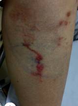 |
| Larvae migrate with vasodilation in the thorax. | The worm larvae move the arm shoulder clearly | Worm larvae migrate anteriorly to the lower leg. |
 |  |  |
| Worm larvae migrate in the back of the calf | Worm larvae migrate on the ankle | Larvae migrate in the abdominal skin and cause secondary infection. |
 |  |  |
| The worm larvae migrate on both sides of the chest area. | Worm larvae moving on the forehead skin of female patient | Larvae migrating with bleeding in the feet |
 |  | 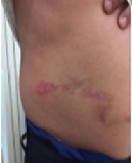 |
| Larvae of worms move in the palms, causing blisters. | Worm larvae migrate on the abdomen around the navel | The worm larvae migrate on a flank‐ventral region. |
 | 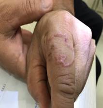 |  |
| The worm larvae move up the entire foot. | Worm larvae migrate on the skin of the finger | Larvae migrate anteriorly to the tibia. |
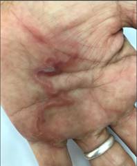 |  |  |
| Worm larvae moving in the palm of the hand | Migratory worms in fingers and back of hand | Worm larvae migrate throughout the palm |
 |  |  |
| Worm larvae move under one shoulder | Larvae migrating in the eye through ophthalmoscopy | Migrating worm larvae cause eye hemorrhages |
 |  |  |
| Migrating larvae cause edema around the eye muscles, creating the typical Zangte’s edema sign. | The worm larvae move and crawl out of small holes. | |

- Regarding the diagnosis of the case: The migrating larvae or the crawling rash usually appear about 1‐5 days after they enter or penetrate the skin, but the incubation period can last for more than 30 days. The typical clinical picture is a serpiginous, erythematous track and is accompanied by itching and mild swelling over the migration trail;
- Common locations are the feet, buttocks, back of the calves, lower limbs, neck, face, cheeks, forehead, knees, ribs, eyes and central nervous system, … or more precisely, locations where the skin comes into contact or is exposed to contaminated soil and surfaces (refer to Dermatologic Conditions);
- CLM is diagnosed clinically based on a history of potential exposure to environmental contaminants and skin lesions. Skin biopsy is not usually recommended. Clinicians can obtain sufficient information for diagnosis through the US Centers for Disease Control and Prevention, including the Division of Parasitic Diseases and Malaria. DPDx laboratory (dpdx@cdc.gov), or references from Parasitic Diseases Hotline for Healthcare Providers ‐ parasites@cdc.gov;
- In terms of differential diagnosis with CLMs: Attention should be paid to scabies and superficial fungal infections. (superficial fungal infections) or deep fungal infections based on clinical and laboratory findings. Characteristics of CLM is quite typical and rarely missed. This type of dermatitis requires a differential diagnosis from other parasitic diseases, subcutaneous nodules or granulomas caused by other parasites and maggots, but also from more common and simple pathologies such as contact dermatitis, atopic dermatitis, superficial urticaria, other dermatitis or pyoderma. Other differential diagnoses include urticaria picta, burrowing scabies, photodermatitis, erythema chronicum migrans and insect or jellyfish stings.
Handling attitude:
+ CLM is usually self‐limiting, migrating larvae usually die after 5‐6 weeks;
+ Albendazole is an effective drug. Some other drugs can be substituted such as ivermectin or thiabendazole. The drug can be taken orally and applied topically;
+ Symptomatic treatment can help relieve itching and reduce bacterial superinfection;
+ Although the infection may resolve spontaneously after a few weeks, secondary infections and symptoms need to be treated;
+ Treatment should include both oral and topical antihelminthic therapy. Oral medications are often preferred by patients because they are generally well tolerated and easier to use and more readily available than topical creams. ivermectin and albendazole are effective drugs of choice. Type ofalbendazole fat form 10% and thiabendazole 15% dạng lỏng hay kem có thể dùng thay thể.
Source
- 1.Neafie RC, Meyers WM. Cutaneous larva migrans. In: Strickland GT ed. Hunters Tropical Medicine and Emerging Infectious Diseases, 8th edn. Philadelphia: Sounders 2000: 797‐799.
- 2.Nash T. Visceral larva migrans and other unusual helminth infections. In: Mandell G, Douglas R, Bennett J, eds. Principle and Practice of Infectious diseases, 3rd edn. New York, NY: Churchill Livingstone 1990: 2340.
- 3.Crocker HR. Diseases of the Skin, 2nd edn. Philadelphia 1893: 926‐927.
- 4.Beaver CP, Jung CR, Cupp EN. Strongylida: Hookworms and other bursate nematodes. In: Clinical Parasitology, 9th edn, Philadelphia: Lea & Febiger 1984: 269‐301.
- 5.Davies HD, Sakuls P Keystone JS. Creeping eruption. A review of clinical presentation and management of 60 cases presenting to a tropical disease unit. Arch Dermatol 1993; 129:528‐529.
- 6.Roberts LS, Janovy J. Foundations of Parasitology. 6th edn. San Francisco: McGraw Hill 2000: 405‐417.
- 7.Gilman RH. Intestinal nematodes that migrate through skin and lung. In: Strickland GT ed. Hunter’s Tropical Medicine and Emerging Infectious Disease, 8th edn. Philadelphia: Sounders 2000: 730‐735.
- 8.Blackwell V, Vega‐Lopez F. Cutaneous larva migrans: Clinical features and management of 44 cases presenting in the returning traveler. Br J Dermatol 2001; 145: 434‐437.
- 9.Jelink T, Maiwald H, Nothdruft HD, et al. Cutaneous larva migrans in travelers: synopsis of histories, symptoms and treatment of 98 patients. Clin Infect Dis 1994; 19:1062‐1064.
- 10.Canizares O. Clinical Tropical Dermatology, Boston: Blackwell Scientific Publications Inc: 1975;210‐21
- 11.Hotez PJ, Narasimhan S, Haggerty J, et al. Hyaluronidase from infective Ancylostoma hookworm larvae and its possible function as a virulence factor in tissue invasion and in cutaneous larva migrans. Infect Immun 1992; 609(3): 1018‐1023.
- 12.Katz R, Ziegler J, Blank H. The natural course of creeping eruption and treatment with thiabendazole. Arch Dermatol 1965; 91:420‐424.
- 13.Bryceson ADM, Hay RJ. Parasitic worms and protozoa. In: Champion RH, Burton JL, Burns DA, et al eds. Rook/ Wilkinson / Ebling Textbook of Dermatology, 6th edn. Vol 2, Oxford: Blackwell Sciences 1999: 971‐972.
- 14.Gilles HM. Soil transmitted Helminths (geohelminths). Cutaneous larva migrans. In: Cook GC ed. Monsor s Tropical Diseases. London: WB Sounders 1996:1392‐1394.
- 15.Gutierrez Y. Diagnostic Pathology of Parasitic Infections with Clinical Correlations, Second edn, New York: Oxford University Press 2000: 343‐353.
- 16.Arthur RP Shelly WB. Larva currens. A distinctive variant of cutaneous larva migrans due to Strongyloides stercoralis. Arch Dermatol 1958; 78:186‐190
- 17.Stone OJ, Newell GB, Mullins JF. Cutaneous Strongyloidiasis: larva currens. Arch Dermatol 1972; 106: 734‐736.
- 18.Roeckel IE, Lyons ET. Cutaneous larva migrans, an occupational disease. Ann Clin Lab Sci 1977; 7:405‐410.
- 19.Burks JW, Jung RC. A new type of water dermatitis in Louisiana. South Med J 1960; 53:716‐719.
- 20.Bhaibulaya M, Chareonlarp P. Creeping eruption caused by Gnathostoma spinigerum. Southeast Asian J Trop Med Public Health 1983; 14:266‐268.
- 21.Daengsvang S, Sermswatsri B, Youngyi P, et al. Penetration of skin by Gnathostoma spinigerum larvae. Ann Trop Med Parasitol 1970; 64:399‐402.
- 22.Pinkus H, Fan J, De Giusti D. Creeping eruption due to Gnathostoma spinigerum in a Taiwanese patient. Int J Dermatol 1981; 20: 46‐49.
- 23.Austman KJ. Creeping eruption: report of first case from Manitoba. JAMA 1926; 78: 186‐190.
- 24.Burtland RJA, Coulson IH. Pulmonary eosinophilia associated with cutaneous la+‐9 migrans. Thorax 1985; 40:76‐77.
- 25.Gnuill MA, Odom RB. Larva migrons complicated by Loeffler’s syndrome. Arch Dermatol 1978; 114:1525‐1526.
- 26.Nabath RP Lowlor PP Nematode (Ancylostoma) in the cornea. A case report. Am J Ophthalmol 1965; 59: 486‐490.
- 27.Little MD, Halsey NA, Cline BL, et al. Ancylostoma larva in a muscle fiber of man following Cutaneous larva migrans. Am J Trop Med Hyg 1983; 32: 1285‐1288.
- 28.Convit J. Protozoan diseases of the skin. In; Elder D, Elenitsas R, Jaworsky C, et al eds. Lever’s Histopothology of Skin, 8th edn, Philadelphia: Lippincott Raven 1997: 1392‐1394.
- 29.Nichols RL. Etiology of visceral migrans. II. Comparative larval morphology of Ascaris lumbricoides, Ancylostoma caninum. J Parasitol 1956; 42: 363‐369.
- 30.Elsner E, Thewes M, Worret WI. Cutaneous larva migrans detected by epiluminescent microscopy. Acta Derm Venereal (Stockh) 1997; 77: 487‐488.
- 31.Rizzitelli G, Scarabelli G, Veraldi S. Albendazole: a new therapeutic regimen in cutaneous larva migrans. Int J Dermatol 1997; 36:700‐703.
- 32.Albanese G, Venturi C, Galbiati G. Treatment of larva migrans cutanea (creeping eruption): a comparison between albendazole and traditional therapy. Int J Dermatol 2001; 40:67‐71.
- 33.Pouluzzi P, MagatonRizzi G, Mattighello P. Larva migrans: report of three cases. Therapeutic advice. J EurAcad Dermatol Venereol 1996; 6: 89‐91.
- 34.Elliot DL, Tolle SW, Goldberg L, et al. Pet associated illness. N Eng J Med 1985; 31: 985‐995.
- 35.Whiting D. The successful treatment of creeping eruption with topical thiabendazole. S Afr Med J 1976; 50: 253‐254.
- 36.Goldsmith J, Froese E. A note on cutaneous larva migrans and its treatment with topical thiabendazole. S Afr Med J 1976; 50: 253‐255
- 37.Stone O, Mullins J. Thiabendozole effectiveness in the creeping eruption. Arch Dermatol 1965; 91: 427‐429.
- 38.Coulaud JP Binet D, Voyer C, et al. Treatment of cutaneous larva migrans with albendazole. Bull Soc Fathol Exot 1982; 75:534‐537.
- 39.Williams HC, Monk B. Creeping eruption stopped in its tracks by albendazole. Clin Exp Dermatol 1989; 14:355‐356.
- 40.Jones SK, Reynolds NJ, Oliwiecki S, et al. Oral albendazole for the treatment of cutaneous larvae migrans. Br J Dermatol 1990; 122:99‐101.
- 41.Veraldi S, Rizzitelli G. Effectiveness of new therapeutic regimen with albendazole in cutaneous larva migrans. EurJ Dermatol 1999; 9; 352‐353.
- 42.Thappa DM, Karthikeyan K. Larva migrans eruption in an infant (Images in Clinical Practice). Indian Pediatr 2002 (in press).
- 43.Caumes E, Carrieri J, Datry A, et al. A randomized trial of ivermectin versus albendazole for the treatment of cutaneous larva migrans. Am J Trop Med Hyg 1993; 49:641‐644.
- 44.Caumes E, Datry A, Paris L, et al. Efficacy of ivermectin in the therapy of cutaneous larva migrans. Arch Dermatol 1992; 128:994
- 45.Edeglass JW, Douglass MC, Stieffer R, et al. Cutaneous larva migrons in the northern climates: a souvenir of your dream vacation. J Am Acad Dermatol 1982; 7:353‐358.
Dr. Huynh Hong Quang
IMPE Quy Nhon


