

The incubation period of tapeworms can range from 20 days to 3 years. The clinical features of tapeworm disease depend on which organ or tissue in the human body is affected. Subcutaneous tissues are most commonly affected by this parasite, but viscera and eyeballs are dangerous if involved and sometimes tapeworms can cause disease in the brain (although rare).

Continued Part 1
Biological cycle & development of tapeworm disease
Adult worms lay eggs that fall into the water, and the eggs are eaten by plankton and crustaceans (first intermediate hosts). These plankton and crustaceans are then eaten by frogs, toads, snakes or birds (second intermediate hosts). When parasitizing in the intermediate host, the larvae are in the form of worms (plerocercoids) about a few centimeters long, ivory‐white, unsegmented, without a head (scolex) in the front, only a pseudo‐sucker.
Humans can become secondary hosts in cases such as drinking water contaminated with pathogens, having plankton and crustaceans infected with tapeworms; eating raw, undercooked frogs, toads, snakes, birds. From here, tapeworm larvae penetrate the digestive tract, burrow into the stomach and intestines and form tumors there. In addition, some backward living habits in rural areas of Vietnam, such as people have the notion that if red eye is treated with raw frog meat, which is cool and cold, and applied to the eyes to cool down, the disease will be cured. This unscientific practice creates favorable conditions for tapeworm larvae to penetrate the skin and eyes, causing eye tumors, which can cause blindness. In some other less common cases, humans can also be infected with tapeworm larvae by washing their faces with water containing plankton and crustaceans infected with tapeworm larvae.
When the second host is invaded and parasitized by the larvae, the disease will occur. The symptoms of the disease depend on where the worm larvae parasitize. If the parasite is in the eye, it will cause pain, watery eyes, inflammation of the cornea, eyelids, and eyelids. If the parasite is in the skin, it will cause itching, rash, and infiltration around the parasite site. Sometimes the patient feels like the larvae are moving.
The life cycle is similar to that of D. latum, but the animals that serve as intermediate hosts and paratenics include humans, mammals, birds, crustaceans, and amphibians. The definitive host is infected by ingestion of asexually encapsulated plerocercariae that are present in the intermediate/paratenic host, which adhere to the intestinal wall and mature. In rare cases, these encapsulated larvae penetrate various tissues, where they remain parasitic without further development. It is also argued that in rarer cases, development to the adult stage in humans has occurred in some reported cases (Miyazaki et al., 1991).
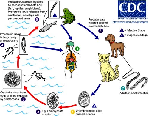
Adult Spirometra tapeworms live in the small intestine of their definitive hosts, dogs, cats, raccoons, and other mammals, for up to nine years, where they lay numerous eggs. When these hosts defecate, the unfertilized/embryoed eggs pass out of the body in the feces and hatch when they fall into fresh water. The eggs are swallowed by crustaceans (molluscs of the genus Cyclops) which are the first intermediate hosts. In copepods, the eggs develop into hook‐tailed larvae (procercoid larvae) that live in body cavities.
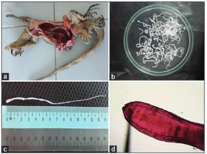
The second intermediate hosts include fish, reptiles and amphibians that feed on crustaceans. The larvae penetrate the intestinal wall of the second intermediate hosts, where they become plerocercoid larvae and proliferate in the subcutaneous tissue and muscle. The second intermediate hosts are eaten by carnivorous definitive hosts such as dogs and the cycle begins again. Humans are accidental hosts in the developmental cycle, becoming infected with asexual plerocercoid larvae through exposure to or ingestion of infected primary and secondary intermediate hosts. The larvae migrate to the subcutaneous tissues of humans. However, no development occurs and there is no ability to transmit the disease. In S. proliferum, multiple larvae can in some cases proliferate throughout the subcutaneous tissue of the body.
The biological cycle and pathogenesis of tapeworms in humans can be summarized as follows: Adult tapeworms of the genus Spirometra live in the intestines of dogs and cat ⑦. Eggs hatch in the feces① and embryonate in the environment under suitable conditions②. Eggs are laid in water and then lyse to form coracidia larvae ③, which are eaten by copepods. The coracidia larvae then develop into procercoid larvae and lodge in the intermediate host, a copepod ④. The second intermediate host includes some fish, crustaceans and amphibians that have ingested infected copepods and contracted these procercoid larvae. The procercoid larvae then develop into adult procercoid larvae inside this second intermediate host for a period of time ⑤. The cycle is completed when a dog or cat eats an infected second intermediate host ⑥. Humans cannot serve as a definitive host for Spirometra spp., but serve as a paratenic or second intermediate host ⑧and develop cysticercosis. Humans acquire cysticercosis by drinking water contaminated with infected copepods or by ingesting undercooked eels.
Tapeworms can live up to 20 years in a human host. The parasite is transmitted to humans in three different ways:
First, humans can become infected by drinking water contaminated with copepods. Second, humans can become infected by eating the raw flesh of one of the intermediate hosts, such as frogs or snakes. For example, people eat live snakes or tadpoles for medicinal purposes in some Asian cultures; if the snakes or tadpoles are infected, the larvae can infect humans.
Third, humans can be infected with tapeworms by poultices prepared from second intermediate hosts applied to open wounds, or acquired for medical or religious reasons. If these poultices are contaminated with plerocercoid larvae, humans can be infected.
According to Zunt et al., human infection most often occurs after ingestion of infected raw snake, frog, or pig meat, although contact with infected meat from intermediate hosts can also cause parasitic infection.
Clinical symptoms
The incubation period of tapeworm can range from 20 days to 3 years.
The clinical features of tapeworm disease depend on which organ or tissue in the human body is affected. Subcutaneous tissue is most commonly affected by this parasite, but viscera and eyeballs are at risk, and occasionally the tapeworm can cause disease in the brain (although this is rare). The early ATDC stage in the development process is usually asymptomatic, but when it reaches its final location and begins to grow, it begins to cause symptoms, creating inflammation and pain in the surrounding tissues.
The clinical features of echinococcosis usually occur after the larvae have migrated to subcutaneous sites. The larval site is usually a tissue or muscle of the chest, abdominal wall, limbs, or scrotum. Although other sites such as the eye, brain, urinary tract, pleura, pericardium, or spinal cord may also be involved. The early stages of the disease in humans are usually asymptomatic, but when typical, the worms can cause inflammation and pain in the tissues surrounding the subcutaneous site where they are developing. Occasionally, S. proliferum can cause proliferative lesions in infected tissues, characterized by the presence of multiple plerocercoids in the same site.
Clinical forms
‐ Ocular sparganosis
This is the most typical form of echinococcosis, the ocular disease has a distinctive appearance and clinical characteristics of echinococcosis. Early symptoms include eye pain, epiphora and optosis. Other symptoms include periorbital edema and swelling resembling the “Romana” sign of exposed corneal ulcer. The most common symptom is a mass lesion in the eye, which, if left untreated, can lead to blindness.
When the larvae enter the eye, it creates a painful reaction, especially edema around the eyeball, which can lead to blindness if not intervened promptly because the parasite moves into the conjunctiva and into the eye socket. In the tissues of the eye socket, the larvae remain at the posterior pole, causing an inflammatory reaction, leading to bulging eyes and impaired vision, some cases with corneal ulcers. In general, this form often causes eye pain, soreness, irritation, corneal ulcers, watery eyes, and significant swelling of the eyelids.

The eye manifestations are usually after 1 week with the manifestation of swollen eyes, redness, reduced vision and sometimes watery eyes, which can easily be confused with other eye diseases, especially corneal disorders, causing doctors to easily diagnose late and not treat promptly if they do not think about it, even patients arbitrarily use medicine to treat themselves with some eye drops. Only when going to a specialist for examination and testing can we discover that the disease is caused by the larvae of the tapeworm S. erinacei. The disease can cause damage and infection to spread to the eyeball and eye socket, and if there is a secondary infection, the disease becomes more severe.
‐ Cerebral sparganosis
This form of the disease is characterized by partial seizures, confusion, weakness, headache, memory loss, coma, fever, paresthesia, motor weakness, and other central nervous system symptoms. This form usually involves one cerebral hemisphere, especially the frontoparietal lobes, in some cases extending to the cerebellum. The disease can cause cerebral hemorrhage.
Multiple discrete subcutaneous nodules may appear and disappear over time. The nodules are often itchy, swollen, red, and mobile, often accompanied by painful edema. Seizures, convulsions, hemiparesis, and headache are also common symptoms of the disease, especially cerebral tapeworms and increased BCAT are also common paraclinical signs in such cases.
The eye manifestations are usually after 1 week with the manifestation of swollen eyes, redness, reduced vision and sometimes watery eyes, which can easily be confused with other eye diseases, especially corneal disorders, causing doctors to easily diagnose late and not treat promptly if they do not think about it, even patients arbitrarily use medicine to treat themselves with some eye drops. Only when going to a specialist for examination and testing can we discover that the disease is caused by the larvae of the tapeworm S. erinacei. The disease can cause damage and infection to spread to the eyeball and eye socket, and if there is a secondary infection, the disease becomes more severe.
‐ Cerebral sparganosis
This form of the disease is characterized by partial seizures, confusion, weakness, headache, memory loss, coma, fever, paresthesia, motor weakness, and other central nervous system symptoms. This form usually involves one cerebral hemisphere, especially the frontoparietal lobes, in some cases extending to the cerebellum. The disease can cause cerebral hemorrhage.
Multiple discrete subcutaneous nodules may appear and disappear over time. The nodules are often itchy, swollen, red, and mobile, often accompanied by painful edema. Seizures, convulsions, hemiparesis, and headache are also common symptoms of the disease, especially cerebral tapeworms and increased eosinophilia are also common paraclinical signs in such cases.


In one case of brain infection with S. erinaceieuropaei, a man presented with headaches, seizures, memory loss, and a strange sense of smell. MRI showed a cluster of rings, initially in the right medial temporal lobe but then moving over time to other areas of the
The cause was not determined for four years until the disease was discovered, and a tissue biopsy finally revealed a 1 cm long worm found and removed. The patient continued to suffer from the same symptoms.
REFERENCES
1. Meghana V. Chougule, Aniruddha Mohite, Vijay P. Joshi, Amit Agrawal (2023). Cerebral sparganosis: Rare parasitic infection of the brain. Egyptian Journal of Neurosurgery volume 38, Article number: 72 (2023)
2. Hu, D.D., Cui, J., Xiao, D., Wang, L., Liu, L.N., Liu, R.D., Zhang, J.Z. & Wang, Z.Q. (2014) Identification of early diagnostic antigens from Spirometra erinaceieuropaei sparganum soluble proteins using immunoproteomics. Southeast Asian Journal of Tropical Medicine and Public Health45, 576‐583.
3. Wael M Lotfy (2020). Neglected rare human parasitic infections: Part I: Sparganosis.Article in Parasitologists United Journal · April 2020
4. Eric Yang, Jonathan Lee, Vishal Patel (2022). Diagnosis and management of cerebral sparganosis: An uncommon parasitic infection of the brain. Radiol Case Rep. 2022 Jun; 17(6): 1874‐1880.
5. Jialing Hu, Kaili Liao, Xiaojin Feng, Danling Jiang, Hailin Liu, Qingcui Zheng, Hai Qiu, Fuzhou Hua, Guohai Xu & Chunhua Xu (2021). Surgical treatment of a patient with live intracranial sparganosis for 17 year. BMC Infectious Diseases volume 22, Article number: 353 (2022)
6. Taisei Kikuchi, Haruhiko Maruyama (2020). Human proliferative sparganosis update. Parasitology International, Volume 75, April 2020, 102036.
7. Jeong‐Geun Kim, Chun‐Seob Ahn, Woon‐Mok Sohn, Yukifumi Nawa, Yoon Kong (2018). Human Sparganosis in Korea. Journal of Korean Medical Science 2018; Infectious Diseases, Microbiology & Parasitology, 33(44): e273.
8. Zhang P, Zou Y, Yu F‐X, Wang Z, Lv H, Liu X‐H, et al. (2019) Follow‐up study of high‐dose praziquantel therapy for cerebral sparganosis. PLoS Negl Trop Dis 13(1): e0007018.
9. M Teresa Galán‐Puchades (2019). Diagnosis and treatment of human sparganosis. The LNCET, Infectious Diseases, Correspondence| Volume 19, Issue 5, p465, May 2019
10. Veronika Muigg, Marie-Therese Ruf, Stefan Schwarzkopf, Simon Huang, Natalja Denisjuk, Anna Stürmann, Michael Ritzler, Rahel Wampfler, Sven Poppert, Andreas Neumayr (2019). Case report: Human subcutaneous sparganosis in a Thai migrant. Am J Trop Med Hyg. 2019 Nov; 101(5): 1170‐1173.
11. Jian‐Feng Fan, Sheng Huang, Jing Li, Ren‐Jun Peng, He Huang, Xi‐Ping Ding, Li‐Ping Jiang, Jian Xi (2021). A human case of lumbosacral canal sparganosis in China. Parasites Hosts Dis. 2021;59(6):635‐638. Published online December 22, 2021
12. Hong D., Xie H., Wan H., An N., Xu C., Zhang J (2018). Efficacy comparison between long‐term high‐dose praziquantel and surgical therapy for cerebral sparganosis: A multicenter retrospective cohort study. PLoS Negl. Trop. Dis. 2018;12(10).
13. Greninger A.L., Glaser C.A. In: Swaiman’s Pediatric Neurology. 6th ed. Swaiman K.F., Ashwal S., Ferriero D.M, Schor N.F, Finkel R.S, Gropman A.L, Pearl P.L, Shevell M.I, editors. Elsevier; 2017. 116 ‐ Fungal, Rickettsial, and parasitic diseases of the nervous system; pp. 907‐917.
14. Liu Q., Li M.‐W., Wang Z.‐D., Zhao G.‐H., Zhu X.‐Q. Human sparganosis, a neglected food borne zoonosis. Lancet Infect. Dis. 2015;15(10):1226‐1235.
15. Ke‐Bin Cheng, Bei‐Lan Gao, Jin‐Ming Liu, Jin‐Fu Xu (2014). Pulmonary sparganosis mansoni: A case report from a non‐endemic region. Journal of Thoracic Diseases.http://dx.doi.org/
16. Deng L., Xiong P., Qian S. Diagnosis and stereotactic aspiration treatment of cerebral sparganosis: summary of 11 cases: Clinical article. J. Neurosurg. 2011;114(5):1421‐1425.
17. Viroj Wiwanitkit (2005). A review of human sparganosis in Thailand. International Journal of Infectious Diseases (2005) 9, 312‐316
18. Kołodziej‐Sobocińska M., Miniuk M. Sparganosis: Neglected zoonosis and its reservoir in wildlife. Med. Weter. 2018;74(4):219‐222.
19. Hughes A.J., Biggs B.A. Parasitic worms of the central nervous system: An Australian perspective. Intern. Med. J. 2002;32(11):541‐553.
20. Moon W.K., Chang K.H., Cho S.Y., Han M.H., Cha S.H., Chi J.G., et al. Cerebral sparganosis: MR imaging versus CT features. Radiology. 1993;188(3):751‐757.
21. Song T., Wang W.‐S., Zhou B.‐R., Mai W.‐W., Li Z.‐Z., Guo H.‐C., et al. CT and MR characteristics of cerebral sparganosis. AJNR Am. J. Neuroradiol. 2007;28(9):1700‐1705.
22. Eberhard M.L., Thiele E.A., Yembo G.E., Yibi M.S., Cama V.A., Ruiz‐Tiben E. Thirty‐seven human cases of sparganosis from Ethiopia and South Sudan caused by Spirometra Spp. Am. J. Trop. Med. Hyg. 2015;93(2):350‐355.
23. Cheng K.‐B., Gao B.‐L., Liu J.‐M., Xu J.‐F. Pulmonary sparganosis mansoni: a case report from a non‐endemic region. J. Thorac. Dis. 2014;6(6):E120‐E124.
24. Wiwanitkit V. A review of human sparganosis in Thailand. Int. J. Infect. Dis. IJID Off. Publ. Int. Soc. Infect. Dis. 2005;9(6):312‐316.
25. Khurana S., Appannanavar S., Bhatti H.S., Verma S. Sparganosis of liver: a rare entity and review of literature. BMJ Case Rep. 2012;2012
26. Hong S.J., Kim Y.M., Seo M., Kim K.S. Breast and scrotal sparganosis: sonographic findings and pathologic correlation. J. Ultrasound Med. Off. J. Am. Inst. Ultrasound Med. 2010;29(11):1627‐1633.
27. Park W.H., Shin T.Y., Yoon S.M., Park S.‐H., Kang Y.J., Kim D.K., et al. A case report of testicular sparganosis misdiagnosed as testicular tumor. J. Korean Med. Sci. 2014;29(7):1018‐1020.
28. Kim J.‐G., Ahn C.‐S., Sohn W.‐M., Nawa Y., Kong Y. Human sparganosis in Korea. J. Korean Med. Sci. 2018;33(44):e273.
29. Fabiani S., Bruschi F. Neurocysticercosis in Europe: still a public health concern not only for imported cases. Acta Trop. 2013;128(1):18‐26.
30.Del Brutto O.H., Garcia H.H. Neurocysticercosis. Handb. Clin. Neurol. 2013;114:313‐325.
31.Bouteille B. Epidemiology of cysticercosis and neurocysticercosis. Med. Sante Trop. 2014;24(4):367‐374.
32. Garcia H.H., Nash T.E., Del Brutto O.H. Clinical symptoms, diagnosis, and treatment of neurocysticercosis. Lancet Neurol. 2014;13(12):1202‐1215.
33. Chang K.H., Cho S.Y., Chi J.G., Kim W.S., Han M.C., Kim C.W., et al. Cerebral sparganosis: CT characteristics. Radiology. 1987;165(2):505‐510.
34. Chang K.H., Chi J.G., Cho S.Y., Han M.H., Han D.H., Han M.C. Cerebral sparganosis: analysis of 34 cases with emphasis on CT features. Neuroradiology. 1992;34(1):1
35. Liao H., Li D., Zhou B., Liu J., Li Y., Liu H., et al. Imaging characteristics of cerebral sparganosis with live worms. J. Neuroradiol. J. Neuroradiol. 2016;43(6):378‐383.
36. Meng Y., Kuang Z., Liao L., Ma Y., Wang X. Case report: morphologic and genetic identification of cerebral sparganosis. Am. J. Trop. Med. Hyg. 2019;101(5):1174‐1176.
37. Zhang P., Zou Y., Yu F.‐X., Wang Z., Lv H., Liu X.‐H., et al. Follow‐up study of high‐dose praziquantel therapy for cerebral sparganosis. PLoS Negl. Trop. Dis. 2019;13(1)
38. Li H.‐X., Luan S.‐H., Guo W., Hua L.‐Y., Zhu H.‐D., Deng J.‐J., et al. Sparganosis of the brain: a case report and brief review. Neuroimmunol. Neuroinflammation. 2017;4(1):238‐242.
39. Zhu Y., Ye L., Ding X., Wu J., Chen Y. Cerebral sparganosis presenting with atypical postcontrast magnetic resonance imaging findings: a case report and literature review. BMC Infect. Dis. 2019;19:748.
40. Torres J.R., Noya O.O., Noya B.A., Mouliniere R., Martinez E. Treatment of proliferative sparganosis with mebendazole and praziquantel. Trans. R. Soc. Trop. Med. Hyg. 1981;75(6):846‐847. doi: 10.1016/0035‐9203(81)90428‐4.
41.Boonyasiri A., Cheunsuchon P., Suputtamongkol Y., Yamasaki H (2014). Nine human sparganosis cases in Thailand with molecular identification of causative parasite species. Am. J. Trop. Med. Hyg., 91: 389‐393, 2014.
42.Quan Liu, Ming‐Wei Li, Ze‐Dong Wang, Guang‐Hui Zhao, Xing‐Quan Zhu. Human sparganosis, a neglected food borne zoonosis. The Lancet Infectious Diseases.2015; 15(10): 1226.
43.R. S. Petrigh, N. P. Scioscia, G. M. Denegri, M. H. Fugassa. Research Note. Cox‐1 gene sequence of Spirometra in Pampas foxes from Argentina. Helminthologia.2015;
44. Wanchai Maleewong, Pewpan M. Intapan, Adhiratha Boonyasiri (2014). Nine human Sparganosis cases in Thailand with molecular identification of causative parasite species. The American Journal of Tropical Medicine and Hygi.2014; 91(2): 389.
45. Quan Liu, Ming‐Wei Li, Ze‐Dong Wang, Xing‐Quan Zhu (2015). Human sparganosis: A neglected food borne zoonosis. The Lancet Infectious Diseases.2015; 15(10): 1226.
46. Co‐existence of Paragonimus harinasutai and Paragonimus bangkokensis metacercariae in fresh water crab hosts in central Viet Nam with special emphasis on their close phylogenetic relationship Parasitology International 2012 | journal‐article.
47. Rapid and simple identification of human pathogenic heterophyid intestinal fluke metacercariae by PCR‐RFLP Parasitology International 2011 | journal‐article DOI: 10.1016/j.parint.2011.09.004, EID: 2‐s2.0‐82155166402.
48.Neurognathostomiasis, a neglected parasitosis of the central nervous system Emerging Infectious Diseases 2011 | journal‐article DOI: 10.3201/eid1707.101433, EID: 2‐s2.0‐79959904332.
49. Molecular identification of a causative parasite species using formalin‐fixed paraffin embedded (FFPE) tissues of a complicated human pulmonary sparganosis case without decisive clinical diagnosis Parasitology International 2011 | journal‐article
50. Impact of hookworm deworming on anemia and nutritional status among children in Thailand Southeast Asian Journal of Tropical Medicine and Public Health 2011 | journal‐article, EID: 2‐s2.0‐80054958645.
51. Human sparganosis in Thailand: An overview Acta Tropica 2011 | journal‐article DOI: 10.1016/j.actatropica.2011.03.011, EID: 2‐s2.0‐79955612835
52.Human paragonimiasis in Viet Nam: Epidemiological survey and identification of the responsible species by DNA sequencing of eggs in patients’ sputum Parasitology International 2011 | journal‐article.
53.Confirmation of the paraphyletic relationship between families Opisthorchiidae and Heterophyidae using small and large subunit ribosomal DNA sequences Parasitology International, 2011.
54. Serodiagnostic reliability of single‐step enriched low‐molecular weight proteins of Taenia solium metacestode of American and Asian isolates Transactions of the Royal Society of Tropical Medicine and Hygiene 2010.
55. Intrahepatic growth and maturation of Gnathostoma turgidum in the natural definitive opossum host, Didelphis virginiana Parasitology International 2010 | journal‐article.
56. Infection status of the estuarine turtles Kinosternon integrum and Trachemys scripta with Gnathostoma binucleatum in Sinaloa, Mexico Revista Mexicana de Biodiversidad 2010 | journal‐article, EID: 2‐s2.0‐79751486432, Source: Scopus ‐ Elsevier Preferred source
57. Helminthic invasion of the central nervous system: Many roads lead to Rome Parasitology International, 2010 | journal‐article.
58. Haplorchis taichui as a possible etiologic agent of irritable bowel syndrome‐like symptoms Korean Journal of Parasitology, 2010 | journal‐article.
59. Double strand problems: Reverse DNA sequences deposited in the DNA database Korean Journal of Parasitology 2010 | journal‐article
60. A novel sigma‐like glutathione transferase of Taenia solium metacestode International Journal for Parasitology 2010 | journal‐article
61. Short report: Case of gnathostomiasis in Beijing, China American Journal of Tropical Medicine and Hygiene 2009 | journal‐article EID: 2‐s2.0‐59649094206 Source: Scopus ‐ Elsevier
62. Morphological differences and molecular similarities between Paragonimus bangkokensis and P. harinasutai Parasitology Research 2009 | journal‐article Large‐group infection of boar‐hunting dogs with Paragonimus westermani in Miyazaki Prefecture, Japan, with special reference to a case of sudden death due to bilateral pneumothorax Journal of Veterinary Medical Science 2009 | journal‐article
63. Is gnathostoma turgidum an annual parasite of opossums? drastic seasonal changes of infection in didelphis virginiana in Mexico Journal of Parasitology 2009 |Journal article
64. Galβ1‐6Gal, antigenic epitope which accounts for serological cross‐reaction in diagnosis of Echinococcus multilocularis infection Parasite Immunology 2009 | journal‐article
65. Fulminant eosinophilic myocarditis associated with visceral larva migrans caused by Toxocara canis infection Circulation Journal 2009 | journal‐article
66. Lu G G Department of Pathogen Biology,Hainan Medical 2014. Discovery of an endemic area of gnathostoma turgidum infection among opossums, didelphis virginiana, in Mexico Journal of Parasitology 2009 | journal‐article EID: 2‐s2.0‐73949145662 Retrospective epidemiological analysis of sparganosis in mainland China from 1959-2012.
67. Lee Eun Kyoung EK Department of Surgery, Konkuk University Medical Center, Seoul, 2014. Axillary sparganosis which was misunderstood lymph node metastasis during neoadjuvant chemotheraphy in.
68. Bennett Hayley M 2014. The genome of the sparganosis tapeworm Spirometra erinaceieuropaei isolated from the biopsy.
69. Oh Youngmin Y Department of Internal Medicine, College of Medicine, Chungbuk National University, Cheongju 361‐711, 2014. Eosinophilic Pleuritis due to Sparganum: A Case Report.
70. Zhao Yi‐Ming YM Department of Pediatric Surgery, The 2nd Affiliated Hospital & Yuying. Scrotal sparganosis mimicking scrotal teratoma in an infant: a case report and …
71. Wiwanitkit Viroj V Tropical Medicine Unit, Hainan Medical University , Haikou , China ‐ ‐ 2014. Ocular Sparganosis.
72. Hill Ag 2014. A Currumbin Wildlife Sanctuary, 28 Tomewin St, Currumbin, Queensland, 4223, Australia. Acanthocephalan infection and sparganosis in a green tree snake (Dendrelaphis punctulata).
73. Cui Jing J Department of Parasitology, Medical College, Zhengzhou University, 40 Daxue Road, Zhengzhou, 450052, People’s Republic of China, 2014. Molecular characterization of a Spirometra mansoni antigenic polypeptide gene encoding a 28.7 kDa …
74. Ouyang Jinsheng J Department of Respiratory Medicine, the First Affiliated Hospital of Wenzhou Medical University, Wenzhou 325000 2014 Pleural sparganosis: Report of a case and review of the literature.
75. Kim Jeung Il JI Department of Orthopedic Surgery, Medical Research Institute, Pusan National University Hospital, Busan 602‐739, 2014. Intramuscular sparganosis in the gastrocnemius muscle: a case report.
76. Hu Dan Dan, 2014. Identification of early diagnostic antigens from Spirometra erinaceieuropaei sparganum soluble proteins using .
77. Tsuda Hiroyuki H Division of Haematology/Oncology, Kumamoto City Hospital, Kumamoto, 2014. Sparganosis in follicular lymphoma patient.
78. Choi Seung Joon SJ Department of Radiology, Gachon University Gil Hospital, Incheon, 2014. Sparganosis of the breast and lower extremities: Sonographic appearance.
79. Wiwanitkit Viroj V University Editorial Office, Hainan Medical University, Haikou, 2014. Multiple sparganosis.
80. Woldemeskel Moges M University of Georgia, Tifton Veterinary Diagnostic and Investigational Laboratory, Tifton, 2014. Subcutaneous sparganosis, a zoonotic cestodiasis, in two cats.
81. Kołodziej‐Sobocińska Marta M Mammal Research Institute, Polish Academy of Sciences, Waszkiewicza 1, 17‐230 Białowieża, Poland. Electronic address: 2014. The first report of sparganosis (Spirometra sp.) in Eurasian badger (Meles meles).
82.Graham Rondell P D 2013. Sparganosis presenting as a mammographic abnormality.
83.Ho Tsai‐Hsuan, 2013. Ocular sparganosis mimicking an orbital idiopathic inflammatory syndrome.
84. Kim Woo Young , 2013. Response to breast sparganosis.
85. Roh Sang‐Young SY Division of Medical oncology, Seoul St. Mary’s Hospital, Seoul , South 2013. Sparganosis in a patient with diffuse large B cell lymphoma.
86. Schauer F 2013. Travel-acquired subcutaneous Sparganum proliferum infection diagnosed by molecular methods.
87. Hong Daojun 2013. Cerebral sparganosis in mainland Chinese patients.
88. Lee H M 2013. Sparganosis of upper extremity in subcutaneous and intramuscular layers.
89. Hu Dan Dan (2013). Dept. of Parasitology, Medical College, Zhengzhou University, 2013. Immunoproteomic Analysis of the excretory-secretory proteins from Spirometra mansoni sparganum.
90. Mo Zhi‐Shuo 2013. Clinical analysis of 25 sparganosis cases.
91. Chu Shuguang 2013. Magnetic resonance imaging features of pathologically proven cerebral sparganosis.
92. Jong‐Yil Chai, Jae‐Ran Yu, Soon‐Hyung Lee, Suk‐II Kim, Seung‐Yull Cho (1998). Ineffectiveness of praziquantel treatment for human sparganosis (A case report). The Seoul Journal of Medicine, Vol. 29, No.4: 397‐399, Dec.1988.
93.Xiang H, Wang J, Tan D, Xiong Y, Huang P, Shen Y, Xu Y, Gong Z, Hu F, Xu C, Wu J, Liu W, Liu J, Wan H, Hong D and Xie H (2023). The serum IgG antibody level as a biomarker for clinical outcome in patients with cerebral sparganosis after treatment. Front. Immunol. 14:1158635. doi: 10.3389/fimmu.2023.1158635.
94. Mark L. Eberhard, Elizabeth A. Thiele, Gole E. Yembo, Makoy S. Yibi, Vitaliano A. Cama, Ernesto Ruiz‐Tiben (2015). Case report: Thirty‐seven human cases of sparganosis from Ethiopia and South Sudan caused by Spirometra spp. Am. J. Trop. Med. Hyg., 93(2), 2015, pp. 350‐355 doi:10.4269/ajtmh.15‐0236
95. Daojun Hong, Huiqun Xie, Hui Wan, Ning An, Chunhua Xu, Jun Zhang (2018). Efficacy comparison between long‐term high‐dose praziquantel and surgical therapy for cerebral sparganosis: A multicenter retrospective cohort study. PLoS Negl Trop Dis 12(10): e0006918. https://doi.org/10.1371/journal.pntd.0006918
96. Choi MH, Kim HT, Kwak TY, Eom SH, Kim YS, Kwak DH, Kim JH (2012). Praziquantel treatment of an eosinophilic pleuritis patient suspected to be due to Sparganum infection. Infect Chemother. 2012 Dec;44(6):522‐525.
Athor: Ph.D. Dr. Huynh Hong Quang
(Institute of Malariology Parasitology and Entomology Quy Nhon)


