

Löffler syndrome is a respiratory disease associated with eosinophilia and a shadow on X‐ray. It was first described by Löffler in 1932. In 1952, Crofton summarized Löffler syndrome as one of five categories of diseases that cause eosinophilic infiltration of the lungs.
INTRODUCE
Löffler syndrome is a respiratory disease associated with eosinophilia and a shadow on X‐ray. It was first described by Löffler in 1932. In 1952, Crofton summarized Löffler syndrome as one of five categories of diseases that cause eosinophilic infiltration of the lungs.
At the same time, medical literature has also mentioned hLoeffler’s syndrome is a transient respiratory illness associated with eosinophilia and radiographic opacities that Crofton et al proposed to classify as one of the five main types of eosinophilic pneumonias based on basic clinical criteria:
[1] Isolated eosinophilic pneumonia or Loeffler’s syndrome, transient infiltrates;
[2] Persistent eosinophilic pneumonia, or recurrent non‐asthmatic infiltrates;
[3] Pneumonia with increased eosinophilia and asthma, infiltrates and asthma;
[4] Tropical eosinophilic pneumonia, often accompanied by asthmatic syndrome;
[5] Polyarteritis nodosa.
This disease is characterized by some opacities in the lung parenchyma associated with increased eosinophils in the peripheral blood or in the lung tissue due to infiltration.
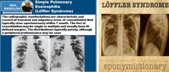

Figure 1. Loeffler syndrome is described in the literature as a transient eosinophilic pneumonia |Source: Löffler syndrome ‐ LITFL ‐ Medical Eponym Library
REASON
The original description of Löffler’s syndrome listed infection with the roundworm A. lumbricoides as the most common cause. However, other parasitic infections and acute drug hypersensitivity reactions have also been implicated as etiologies of pulmonary eosinophilia.
Loeffler’s syndrome can be an acute, often self‐limiting pneumonia associated with eosinophil proliferation in the lungs. It is also commonly caused by allergic reactions to agents such as parasites, molds, or other allergens.
- Parasitic infections: This is the most common cause, especially in tropical areas. Common parasites that cause Loeffler’s syndrome include roundworms, hookworms, and strongyloides;
- Allergic reactions: Allergies to medications, foods, or other irritants can also cause Loeffler’s syndrome;
- Autoimmune diseases: In rare cases, Loeffler syndrome may be associated with autoimmune diseases.
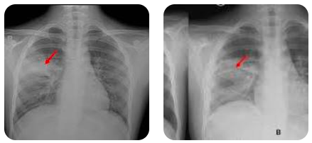
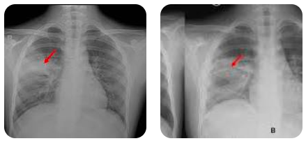
Figure 2. Loeffler syndrome can occur in both sexes and in both adults and young children.
SOME PARASITES CAUSING LOEFFLER’S SYNDROME
1. Ascaris lumbricoidesAscaris lumbricoides:
As the most common cause of Loeffler’s syndrome, roundworms in humans mature from larvae or eggs into adult worms that can grow to over 30 cm in length when they are capable of reproduction. Roundworm infections are common in children in tropical and subtropical regions worldwide, especially in areas with poor sanitation. It is also one of the most common parasitic infections in the world, which often goes unrecognized because most people infected are mild and asymptomatic. However, severe infections can lead to serious symptoms and complications, depending on the parasite load and the tissues or organs affected.
In the lungs, after people swallow eggs worms, They hatch in the small intestine and the larvae migrate through the bloodstream or lymphatic system to the lungs. At this stage, the person may experience signs and symptoms of Loeffler syndrome., similar to asthma or pneumonia, including persistent cough, difficulty breathing, and wheezing. After 10‐14 days in the lungs, the larvae migrate to the throat, where the person coughs them up and often swallows them back into the digestive tract;
In the intestine, the larvae mature into adult worms in the small intestine, and the adult worms re‐locate in the intestine, laying eggs until they die. Symptoms of intestinal invasion are vague abdominal pain, nausea and vomiting, diarrhea or bloody stools. If the patient has a large intestinal worm load, usually children, there will be severe acute abdominal pain, violent vomiting due to intestinal obstruction, and worms appear in the patient’s vomit or stool.
2. Hookworm infection, beak Necator americanus/Ancylostma duodenale
Hookworm is an obligate parasitic roundworm in the small intestine of humans. The habitat of hookworm is in warm climates because the eggs need a moist, warm and shady environment to hatch. The lifespan can be up to 3‐5 years and they can lay from 5,000 to 10,000 eggs per day. The life cycle of this worm begins from unhatched eggs in the soil. After 24‐48 hours, the eggs become embryos and hatch into larvae, molt many times into young worms and can penetrate human skin, pass through the blood vessels and heart, then reach the lungs. Here, it penetrates the alveoli of the lungs and goes up the trachea, is swallowed into the digestive tract, and down to the small intestine. The worms attach to the wall of the small intestine, suck blood, grow into adult worms and begin to reproduce, lay eggs that are excreted in the stool;
During the process of entering the body, some larvae cannot easily pass through the dermis and remain trapped in the skin, causing skin irritation. When reaching the lungs, it causes symptoms such as prolonged cough and difficulty breathing, wheezing ‐ manifestations of Loeffler’s syndrome. When reaching the intestine, the worm can cause a loss of 30 μl of blood per day, causing the patient to have iron deficiency anemia, fatigue in adults and mental retardation and poor development in children. In addition, hookworm infection will often cause abdominal pain, increased after meals, accompanied by diarrhea, bloating and nausea.
3. Strongyloidiasis Strongyloides stercoralis
Is a roundworm belonging to the group of worms transmitted through the soil or through the digestive tract to humans. People are infected mainly through contact with soil infected with free‐living larvae. When the larvae come into contact with the skin, they penetrate the skin, move throughout the body, and eventually reach the small intestine, grow and lay eggs. Unlike other types of soil‐transmitted worms, strongyloides eggs hatch into larvae in the intestine and will be excreted in the stool. Therefore, the larvae that have just been excreted will be able to immediately re‐infect the host by burrowing into the intestinal wall, or penetrating the skin around the anus. It is this mechanism that makes it possible for a person to be autoinfected with strongyloides (autoinfection cycle) throughout life.
However, most people infected with strongyloidiasis have no symptoms. If they do, they usually describe nonspecific discomfort. Some people have abdominal pain, bloating, heartburn, and episodes of diarrhea and constipation. Some people have a dry cough, difficulty breathing, and wheezing and are diagnosed with Loeffler’s syndrome. Others have skin rashes, arthritis, and kidney and heart damage.
4. Infection with roundworm larvae in dogs and cats Toxxocara spp.
Toxocara canis and feline larval infection is a disease belonging to the group of visceral larval migration. Depending on the location of the disease, the level of increased eosinophilia, signs in the eyes or lungs, the terms indicate the movement of the larvae to different tissue and organ locations. In particular, when the worm larvae cause disease in the lungs, the patient also has symptoms similar to the above‐mentioned worm infections such as roundworm, strongyloidiasis and hookworm, diagnosed as Loeffler syndrome.
SOME MEDICINES THAT CAN CAUSE Eosinophilic PNEUMONIA (Eosinophilic Pneumonia)
Various groups of drugs or metals, substances used in medical X‐ray equipment, metal salts, or dust mites… can also cause eosinophilic pneumonia, including:
‐ Acetaminophen, Acetylsalicylic acid, Aluminum, Amiodarone, Amitriptyline, Ampicillin, thuốc ức chế men chuyển angiotensin (ACEI), Azathioprine, Beclomethasone dipropionat, Beryllium, β‐Blockers, Bleomycin, Captopril, Carbamazepin, Chloroquin, Chlorpromazin, Chlorpropamid, Clarithromycin, Clofibrat, Dantrolen, Dapson, Daptomycin, Desipramin, Diclofenac, D‐penicillamine, Ethambutol, Fenbarbamat, Fenbufen, Fludarabine, Glafenine, chất kích thích đại thực bào (MCSFs), Hydrochlorothiazid, Ibuprofen, Imipramine, Indomethacine, Infliximab, Interferon‐alpha, Interleukins, Isoniazid, Levofloxacin, L‐Tryptophan, Maloprim, Mecamylamin, Mephenesin carbamat, Mesalazin, Methotrexate, Methylphenidat, Minocyclin, Montelukast, Naproxen, Nilutamid, Nitrofurantoin, Nomifensin, Oxaliplatin, Para‐aminosalicylic acid, Penicillamin, Penicillin, Phenytoin, Piroxicam, Procarbazine, Progesterone, Prontosil, Propylthiouracil, Pyramethamin, Ranitidine, Salicylazosulfapyridine, Streptomycin
‐ Cocaine (inhalation), contrast agents, Cromolyn (inhalation), Pentamidine (inhalation), house dust mites (inhalation), gold salts, heroin (inhalation), iodine contrast agents, Nickel dust (inhalation), Rapeseed oil, antigens from red spiders, scorpion stings, Sertraline, cigarette smoke, radiation exposure, antibiotics containing the Sulfa‐ group, Sulfasalazine, Sulindac, Tamoxifen, Tetracycline, Thiazide, Tolazamide, Tolfenamic acid, Trazodone, Trichloroethane, Venlafaxine.
PATHOPHYSIOLOGY and SYMPTOMS
Pathophysiology
In the pathophysiological mechanism, Loeffler’s syndrome is associated with the migration of several species of roundworm parasites through the lungs during their biological and developmental cycles in the human body. After ingestion of Ascaris lumbricoides eggs, the larvae hatch in the intestine and penetrate the mesenteric lymphatics and venules to enter the pulmonary circulation. The parasites reside in the pulmonary capillaries and continue the cycle by migrating through the alveolar walls.
Finally, they migrate up the bronchial tree and are swallowed into the esophagus, returning to the intestines and maturing there. This process takes about 10‐16 days after ingestion of Ascaris, Hookworm and Strongyloides eggs. In addition, other parasites, such as Necator americanus, Ancylostoma duodenale and Strongyloides stercoralis, have similar life cycles to Ascaris, with the migration of larval forms through the alveolar walls. Some of these parasites may not be ingested orally but enter the host through the skin.
Recently, Loeffler’s syndrome has also been shown to be associated with the use of certain medications, including antibiotics (ethambutole, isoniazid, nitrofurantoin, penicillin, tetracycline, clarithromycin), anticonvulsants (carbamazepines, phenytoin, valproic acid, ethambutol), anti‐inflammatory and immunosuppressive drugs (aspirin, azathioprine, beclomethasone, methotrexate, naproxen, diclofenac, fenbufen, ibuprofen, phenylbutazone, piroxicam) and other drugs (bleomycin, captopril, chlorpromazine).
In most cases, Loeffler’s syndrome is considered a benign, self‐limiting disease with no significant morbidity and no reported deaths. Because young children are exposed to contaminated soil and exhibit hand‐to‐mouth transmission behaviors more frequently than adults, children have a higher incidence of intestinal helminthiasis and Loeffler’s syndrome. Symptoms of Loeffler’s syndrome usually subside within 3‐4 weeks or shortly after drug withdrawal if drug‐induced pulmonary eosinophilia is suspected.
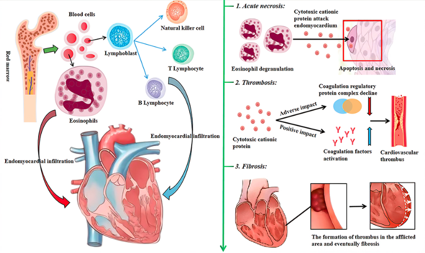
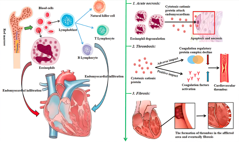
Figure 3. The stages of Loeffler endocarditis should not be confused with endocarditis. Eosinophilic and myeloid lymphocytic endocardial infiltration.
1st Stage Acute necrosis; 2nd Stage thrombus formation; 3rd Stage fibrosis.
Clinical symptoms
Symptoms of Loeffler syndrome are usually mild and transient and may include: Dry or productive cough; shortness of breath, especially with exertion; low‐grade fever; feeling tired, loss of appetite, and rarely chest pain. Loeffler’s syndrome is a rare form of pulmonary eosinophilia, with symptoms that are usually transient, self‐limiting, and benign lasting less than a month (average 6‐12 days). The etiology of Loeffler’s syndrome is thought to be primarily an allergic reaction to the migration of larvae of worms or possibly flukes through the lungs, specifically roundworms, hookworms, and strongyloides.
Symptoms of Loeffler’s syndrome are usually mild, tending to resolve spontaneously after a few days or at most after 2‐3 weeks. A dry cough is the most common symptom. In addition, the patient may have difficulty breathing, wheezing. Some patients may have muscle pain, loss of appetite, and hives. These symptoms usually appear 10‐16 days after ingestion of Ascaris eggs. A similar incubation period has been described in Loeffler’s syndrome associated with infection with N. americanus, A duodenale, or S. stercoralis. Some cases have a history of travel to endemic areas, which would also be a risk factor for exposure to the parasite.
In cases where drug‐related Loeffler syndrome is suspected, evidence of use of the drugs known to cause eosinophilia should be obtained. On examination, a physician may hear crackles or wheezing in a patient with Loeffler syndrome. In contrast, patients with drug‐induced pulmonary eosinophilia often have wheezing on auscultation.
Loeffler syndrome in children is a type of eosinophilic pneumonia similar to community‐acquired pneumonia and asthma caused by roundworm that has been recorded in Vietnam and around the world. For example, here is a case of a pediatric patient Clinical manifestations similar to community‐acquired pneumonia and bronchial asthma. A 5‐year‐old boy presented with symptoms of cough, dyspnea, wheezing, and intermittent fever for three days. He was diagnosed with pneumonia after physical and imaging evaluation and was admitted to a hospital. After discharge, his symptoms improved but persisted for several weeks and he coughed up sputum. When he arrived at the medical facility, his general condition appeared good, but on physical examination, the doctor found that he had rales and coarse sounds, especially in the upper lung region (T). Evaluation of the patient’s peripheral blood test showed increased eosinophilia (from 0.2% to 2.06%‐338/mm3), while IgE was 50 IU/mL.
Chest X‐ray showed patchy consolidation in the pulmonary infiltrate in lung T. Despite antibiotic treatment, the consolidation did not resolve but almost completely disappeared when the patient vomited the parasite. Gross evaluation of the material was confirmed by microbiologists as A. lumbricoides.
|
| 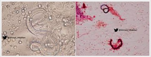  |
| Figure 3. Images of eggs and adult parasites in sputum samples from patients | |
DIAGNOSE
Because the symptoms of Loeffler syndrome are often vague, nonspecific, and transient, diagnosis requires supportive tests, and to diagnose Loeffler syndrome, clinicians will rely on the following information:
(i) Medical history: Ask about symptoms, exposure to allergens or parasites;
(ii) Clinical examination: Cough is the most common symptom in Loeffler’s syndrome, listen to the heart and lungs, check for signs of allergy;
(iii) Blood test shows increased number and proportion of BCAT, with or without increased total IgE in serum. Usually, BCAT slight increase in blood (> 7‐20%); If BCAT accounts for up to 40%, the cause of the disease should be considered to be due to drugs;
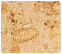
(iv) Stool test may show single cells due to the patient swallowing sputum and passing it out with the stool. Parasites and eggs may be found in the stool for 6‐12 weeks after the primary infection. However, by this time, pulmonary symptoms are self‐limited or have disappeared.;
(v) Analysis of sputum, gastric juice or bronchoalveolar lavage sometimes finds larvae in sputum and gastric aspirate at the time of pulmonary symptoms; Bronchoalveolar lavage: The number of BCAT may be increased;
(vi) Chest X‐ray shows mobile opacities in the lungs. Chest X‐rays may show a opacity that has moved from its previous location 12‐20 days ago. However, the lung lesions seen on the film may completely resolve within 2‐4 weeks. In drug‐induced pulmonary eosinophilia, the radiographic abnormality will completely resolve several weeks after discontinuation of the suspected drug;
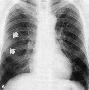 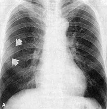 | 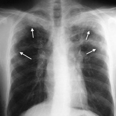  |
| Figure 1. Initial chest radiograph of a 54‐year‐old male patient, showing an opacity (arrow) over the middle lobe of the right lung. | Figure 2. Chest X‐ray shows a moving opacity over time in an untreated patient. |
(vii) CT‐scan provides more detailed images of lung lesions;
(viii) Lung biopsy may be required in some cases to determine the exact cause. Lung lesions show increased eosinophilic infiltration occurring in the bronchi and bronchioles, in the alveoli and interstitium. However, parasitic forms are usually not found in the lungs.
TREATMENT & CASE MANAGEMENT
In many cases, Loeffler’s syndrome resolves spontaneously (2‐3 weeks) without specific treatment. However, if symptoms are severe or complications arise, your doctor may prescribe the following treatments:
- Antihistamines: Reduce allergy symptoms;
- Antibiotics: If there is bacterial superinfection;
- Corticosteroid: Reduce inflammation;
- Antiparasitic drugs: If the cause of the disease is due to parasites. It should be noted that some types of parasites can cause Loeffler’s syndrome such as fascioliasis, fascioliasis, schistosomiasis, T. gondii, tapeworms, filariasis and trichinosis are still possible but rare.
Treatment and monitoring of Loeffler syndrome can be performed on an outpatient basis, without hospitalization. Patients can resume normal physical activity and diet, without special restrictions. Since symptoms in most patients with Loeffler syndrome are self‐limiting, specific treatment is not necessary. If pulmonary eosinophilia is caused by medication, the patient should stop taking the suspected drug. Conversely, if the cause is thought to be parasitic infection, the patient should be prescribed appropriate antihelminthic medication. If the severity of the disease is severe or slow to improve, in both situations, systemic corticosteroid therapy may be highly effective, helping to improve symptoms quickly.
Treatment of helminth infections is only indicated when there is evidence of worm infection in the body. Antiparasitic drugs are the first line of treatment against common human infections, the most common being albendazole, ivermectin, mebendazole. These drugs, when taken for 1‐3 days, kill adult worms. Side effects from these drugs include mild abdominal pain or diarrhea.
In addition, to prevent and avoid reinfection, each person needs to develop good hygiene awareness, wash hands with soap, eat cooked food and drink boiled water, build houses with sanitary waste treatment systems, combined with an educational campaign to eliminate the use of outdated latrines, especially in rural and mountainous areas. For children, parents need to instruct them to wash their hands regularly with soap, not to put their hands in their mouths, to give up the habit of biting their nails, and to go barefoot. For patients who have been diagnosed with increased pulmonary eosinophilia due to drugs, it is necessary to avoid using that drug in the future.
PROGNOSIS
Loeffler syndrome generally has a good prognosis. Most patients recover completely within a few weeks or months. However, in some cases, the disease may recur or progress to chronic bronchial lung disease.
Initially, the most common cause of Loeffler’s syndrome was thought to be Ascaris lumbricoides, but other possible causes of simple eosinophilic pneumonia include acute drug hypersensitivity reactions and parasitic infections. Loeffler’s syndrome is considered a benign, self‐limited disease that is not fatal and leaves few significant sequelae. Symptoms usually resolve within 3 to 4 weeks or less after discontinuation of the causative agent in drug‐induced eosinophilic pneumonia.
REFERENCES
1. Acar A, Oncül O, Cavuşlu S, Okutan O, Kartaloğlu Z. Case report: Löffler syndrome due to Ascaris lumbricoides mimicking acute community‐acquired bacterial pneumonia. Turkiye Parazitol Derg. 2009;33:239‐41.
2. Deveci U, Üstün C, Altınsoy HB, Akay A, Özdiller S, Aydın M. Loeffler syndrome mimicking bronchial asthma and pneumonia in a child: a case report. Turkiye Parazitol Derg. 2013;37:288‐91.
3. Loffler Syndrome 2022. https://emedicine.medscape.com/
4. Oner Ozdemir (2020). Loeffler’s syndrome: A type of eosinophilic pneumonia mimicking community‐acquired pneumonia and asthma that arises from Ascaris lumbricoides in a child. North Clin Istanb 2020 Aug 5;7(5):506‐507.
Dr. Huynh Hong Quang
IMPE Quy Nhon


