

Molecular biology testing (MBT) is a term used to refer to tests that detect biological markers at the molecular level such as nucleic acid fragments, complete genes or microbial genomes.

The SHPT test is a PCR gene synthesis reaction, a leap forward from the success of gene identification and sequencing techniques. This technique aims to determine the cause of the disease more quickly and accurately to serve the treatment process more effectively. In the SHPT test, it is mandatory to use positive and negative controls.
Positive controls are control samples containing a known target DNA/RNA segment. Negative controls are control samples without the target DNA/RNA segment to help control cross‐contamination when performing reactions. The SHPT test uses two main techniques: PCR synthesis and real‐time PCR.
Polymerase Chain Reaction (PCR) in particular and PCR‐based techniques in general have been widely used due to their sensitivity, specificity, and high accuracy in testing results in modern medical fields to detect, diagnose, and analyze the genetics of some specific diseases related to viral, bacterial, parasitic, and fungal agents that we have long faced the challenge of being “difficult or impossible to find with traditional testing techniques”.
This PCR test is especially useful in detecting and diagnosing some agents that are difficult or impossible to culture in routine testing environments such as bacteria ( Mycoplasma spp. , Chlamydia spp.,…), viruses (HIV, Dengue, H5N1, hepatitis B, hepatitis C), parasites (malaria, helminths and protozoans); PCR can help find and detect agents that have been cultured but failed because they were previously dead or had very low density in clinical samples such as “headless/decapitated” purulent meningitis, tuberculosis bacteria that failed to culture; Find and detect drug‐resistant bacteria strains, detect Dengue virus strains that cause dengue fever, identify gene mutations related to cancer, and drug‐resistant gene mutations. In addition to the medical field, the PCR method is also applied in the production of pathogen diagnostic kits in a variety of other fields including: Identifying diseases in humans, veterinary medicine, aquaculture, food microbiology, identification, etc.
1. Overview of PCR technique
Polymerase chain reaction (PCR) is a technique that allows amplification of a gene segment in a test tube based on thermal cycling. Based on the necessary reaction components, the target gene segment will be cloned into millions of identical copies, thereby serving the purpose of further research. This is considered a basic process for the development of a series of gene and genome analysis technologies later.

The PCR technique was developed by American biochemist Kary Mullis based on the idea of creating a process that allows a DNA segment to be artificially replicated through multiple reaction cycles through the catalysis of the DNA polymerase enzyme. This idea was when he wanted to amplify and create multiple copies of a DNA segment without using living organisms such as E. colibacteria or yeast to separate the double‐stranded DNA into two single strands, then use complementary primer pairs to pair with the single strand, and replicate using the DNA polymerase enzyme.
The biggest challenge in genetic analysis in the past was the single, very small targets in a complex, huge genome. The advent of PCR changed all that, making it possible to create large numbers of copies of a desired DNA segment. Interestingly, the DNA polymerase enzyme isolated from E. coli is not heat‐stable and needs to be added after the heating stage of each thermal cycle, making the process slow, expensive and laborious.
This problem was solved when microbiologist Thomas Brock accidentally discovered the heat‐resistant bacteria Thermophilus aquaticus (or Taq) in the hot spring area (>70oC) in Yellowstone (USA) in the early 60s. The discovery of the enzyme Taq polymerase helped PCR become a revolution in molecular genetics, one of the basic techniques applied in all laboratories in the world today.
PCR is now used in a variety of fields, from medicine to paleontology to forensics. One of the most common applications of PCR is in the field of forensic science. Before the advent of PCR, a small sample of DNA may not have been enough to perform an accurate test and draw a definitive conclusion. However, with PCR, researchers can generate enough DNA to identify remains from past accidents or disasters.
Similarly, small DNA samples recovered from crime scenes can be used by PCR to identify or eliminate suspects. In medicine, PCR has become an important tool in cancer research. PCR is used to detect some cancers caused by viruses such as cervical cancer (HPV), skin cancer, and oral cancer. Another important medical application of PCR is in tissue typing to determine the compatibility between donor tissue and patients undergoing organ transplants, to select appropriate treatment for patients, to test for genetic relationships; to identify infectious diseases early and to monitor the spread of infectious diseases in humans. In addition, PCR can identify both fungal and parasitic infections, and to amplify DNA to screen donated blood to prevent infected blood from entering blood banks.
In paleontology, PCR is used to amplify DNA samples that have been present in tissues for hundreds of years. PCR can be used on mummified tissue samples, ancient flora and fauna, or fossils. Thus, PCR can be used to identify and learn about now‐extinct organisms and fossil and evolutionary processes.

PCR is used to help detect and identify harmful bacteria and pathogens in public water supplies; detect degenerative microorganisms where toxic waste and pollution incidents have occurred.
2. Minimum components for PCR reaction
To perform a PCR test, the following components are required: a sample DNA containing a specific purified DNA segment for later cloning; a primer (a DNA primer segment) about a few tens of Kb long to locate the start and end points of the sample DNA segment; a DNA polymerase (an enzyme that synthesizes a copy of the sample DNA segment), which must be a highly heat‐resistant enzyme;
Nucleotides include 4 types A, T, C and G which play a role in forming the structure of the DNA copy; Buffer solution to provide the environment for the DNA polymerase enzyme to work and specialized PCR tubes to mix the PCR reaction solution before putting it into the PCR testing device.
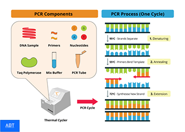
Specifically, thermostable polymerase (Taq polymerase) , this enzyme has an optimal operating temperature of 72 o C; 4 types of nucleotides (dNTP) including dATP/dTTP/dGTP/dCTP are the raw materials to synthesize DNA copies; DNA containing the target sequence (Template), usually in testing, this is DNA obtained from the sample to be tested; Specific primer pairs are oligo‐nucleotide segments about 20 nucleotides long and specifically complementary to the 2 ends of the target sequence to be cloned; Mg 2+ cation (MgCl 2 ) is an important co‐factor for Taq polymerase to work effectively and Tris‐KCl buffer solution (PCR Buffer) provides a favorable environment for the cloning reaction to take place.
The test tube containing the PCR mix will be placed into the incubation chamber of the PCR thermal cycler. The temperature inside the incubation chamber of the PCR machine will change periodically, thanks to which the DNA replication process takes place.
Primer
Primer ‐ also known as primer ‐ is a nucleic acid (or ribonucleic acid ) strand used as a starting segment for the DNA replication process . Most DNA polymerases ( enzymes that catalyze DNA replication) cannot begin synthesizing a new DNA segment without a primer. Because it only attaches nucleotides to the available primer strand according to the principle of complementing the template strand .
In most natural DNA replication processes, the basic primer for DNA synthesis is a short strand of RNA. This RNA is produced by RNA polymerase, which is removed and replaced by DNA by DNA polymerase. Many molecular biological
techniques involving DNA polymerase, such as DNA sequencing and PCR, require primers. Primers for these techniques are usually short ( # 20 bases), synthetic DNA molecules.
The actual structure of the primer begins with a 3′‐hydroxyl nucleoside attached to a controlled ‐pore glass (CPG). The 5′‐hydroxyl nucleoside is capped by dimethoxytrityl (DMT) to prevent the formation of a nucleotide chain. To add a nucleotide, the DMT must be chemically removed, and a nucleotide is added. The 5′‐hydroxyl end of the new nucleotide is blocked by DMT to prevent the addition of one or more nucleotides on a chain.
The cycle is then repeated for each nucleotide using a primer. This is just a simplified description, the actual process is quite complicated. Therefore, most labs do not make primers themselves.

DNA sequencing is used to identify the nucleotides in a DNA strand. The sequencing method, called dideoxy sequencing (also known as the Sanger method ), uses primers as starting markers for the chain reaction. In PCR, primers are used to identify the DNA segments that are amplified by PCR. Primers are usually no longer than 50 nucleotides in length (Since DNA is usually double‐stranded, its length is measured in base pairs.
of single‐stranded DNA is measured in bases or nucleotides), and they match exactly with the beginning and end of the amplified DNA segment. They bindthe DNA template at the beginning and end, where DNA polymerase attaches and begins synthesizing a new DNA strand.
The selection of primer length and its melting temperature is based on the principle : The melting temperature of a primer is a temperature lower than the temperature at which the primer anneals the DNA template and higher than the temperature at which the primer detaches from the DNA template. The melting temperature should increase with the length of the primer. A primer that is too short will anneal or extend ( anneal ) some positions on the long DNA template, resulting in nonspecific copies. In other words, the length of a primer is limited by the temperature at which it melts.

Melting temperatures that are too high, i.e. above 80°C, can cause some problems because DNA polymerase is less active at this temperature. The optimal length of a primer is around 30 ‐ 40 nucleotides with a melting temperature of around 60°C to 75°C. There are several ways to calculate the melting temperature (TM) of a primer.
Digestive primers are sometimes used , which are mixtures of similar, but undefined, primers. They may be appropriate if the same gene is to be amplified from different organisms . Another use for digestive primers is when primer design is based on a protein sequence . Several different codons can code for a single amino acid . Thus the primer sequence corresponding to the amino acid isoleucine might be “ATH”, where A stands for adenine , T for thymine , and H for adenine , thymine , or cytosine . Using digestive primers can reduce the specificity of PCR.
(continued) ‐> Next part: Some molecular biological tests in the study of some infectious diseases in humans (continued)
Dr. Huynh Hong Quang & Dr. Nguyen Thi Lien Hanh
IMPE Quy Nhon


