

Eosinophilic gastritis is also associated with other conditions such as irritable bowel syndrome, collagen vascular disease, bacterial and parasitic infections (including H. pylori), drug injury, and drug sensitivity. Although eosinophilia can be seen in a variety of disease states, eosinophilia may be prominent in allergic and atopic diseases.
DEBATE ON DIAGNOSTIC FRAMEWORK
CDiagnosis requires a combination of clinical history, clinical examination, endoscopy, paraclinical diagnosis and imaging diagnosis to determine the case ( Gut 1990;31:54):
- Gastrointestinal symptoms
- Gastrointestinal tissue biopsy shows increased BCAT
- Exclude previously known causes of increased BCAT.
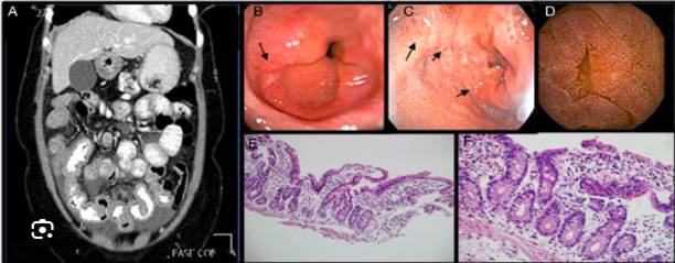
Gastrointestinal endoscopy: The appearance can range from normal to mucosal erythema, background edema, granulomas, ulcers, mucosal thickening, and the formation of fragile white plaques (Clin Rev Allergy Immunol 2016;50:175)
DEBATE ABOUT PARACLINICAL
There is no single test or procedure that can fully diagnose EGIDs
Maintaining a high index of suspicion when performing clinical examination is essential for final diagnosis
Although not specific, the following paraclinical tests can help in diagnosis (Clin Rev Allergy Immunol 2016;50:175):
- Allergy testing to assess which allergens may be triggering the disease and what symptoms may occur This is currently controversial (J Gastroenterol Hepatol 2013;28:1306)
- Increased number of BCAT in peripheral blood (20 ‐ 80% of cases)
- Increased erythrocyte sedimentation rate Increased serum IgE;
- Iron deficiency anemia Hypoalbuminemia;
- Cationic eosinophilic protein content in feces
- Eosinophilic cationic protein and neurotoxin extracted from BCAT (Scand J Gastroenterol 2011;46:1074)
- Increased 2‐macroglobulin (Scand J Gastroenterol 2011;46:1074)
- Fecal examination to remove parasites.
Diagnostic imaging
It is useful but limited
Ultrasound:
- Detect the presence of intestinal wall thickening, abdominal fluid or peritoneal nodules
- Also helps monitor and determine treatment response (Ultraschall Med 2011;32:E57)
CT‐scan:
May show nodules, abnormal folds, and thickening of the gastric and small intestinal mucosa ( J Comput Assist Tomogr 1999;23:417)
Barium enema:
Determine different degrees of gastrointestinal stricture, mucosal abnormalities, and gastric fold thickness ( J Clin Pathol 1986;39:1)
Tc‐99m hexamethyl propylene amine oxime (HMPAO) leukocyte scintigraphy:
- Active inflammatory areas have increased BCAT
- Useful in determining extent of disease and assessing response to treatment (Clin Nucl Med 1997;22:536, Ann Nucl Med 2003;17:601).
PROGNOSIC FACTORS
Unknown due to the rarity of the disease but has been observed to depend on treatment response and Klein classification ( Clin Gastroenterol Hepatol 2011;9:950)
Patients had 3 different disease stages (Clin Gastroenterol Hepatol 2011;9:950):
- Simple outbreak: More related to the serous model
- Recurrence: More related to the body;
- Continuous: More related to the mucosal body.
DIFFERENTIAL DIAGNOSIS
Bacterial or parasitic infection
- Pinworm, hookworms, whipworms and schistosomiasis;
- Toxocara canis can also be a cause of eosinophilic ascites..
H. pylori infection
- Biopsy and silver staining to rule out pylori;
- Case reports of successful treatment of EGIDS with eradication of H.pylori(Gut 2005;54:1822, J Clin Gastroenterol 2008;42:1063)
Irritable bowel disease (Mayo Clin Proc 1997;72:117):
- There may be enteritis with increased peripheral blood eosinophils
- Lack of data on diffuse BCAT increase.
BCAT hyperinfection syndrome
- Rare and rare tissue conditions
- Persistent (> 6 tháng) significant peripheral blood eosinophilia (usually >1.500 BCAT/microliter);
- In the presence of organ damage or organ dysfunction associated with eosinophilic infiltration and lysis of important mediators;
- Most clinical manifestations involve the skin and lungs (J Allergy Clin Immunol 2009;124:1319)
Vasculitis (Churg-Strauss syndrome and polyarteritis nodosa ):
- BCAT infiltration into small blood vessels
- Increased peripheral blood eosinophilia
- Increased inflammatory markers and autoantibodies.
Connective tissue disorders (scleroderma and dermatomyositis):
Intermittent eosinophilia and band‐like mast cell and eosinophil infiltration in the mucosa.
Drug sensitivity:
Gastrointestinal eosinophilia is associated with a very long list of drugs, such as carbamazepine, rifampicin, naproxen, NSAIDs, interferon, azathioprine, enalapril, or gold compounds.
Mastocytosis and Langerhans cell histiocytosi:
- Gastrointestinal eosinophilia is commonly associated with the digestive system by Mastocytosis và Langerhans cell histiocytosi.
- In addition to BCAT, mononuclear cell infiltration is prominent and nodules and granulomas appear
- Mononuclear cells may have morphological characteristics of either mast cells or Langerhans cells as determined by immunohistochemical staining.
DEBATE ON ATTITUDES AND LONG‐TERM TREATMENT
There are currently no published standard treatment guidelines due to the lack of extensive longitudinal studies. Current treatment modalities are based on individual case reports or case series
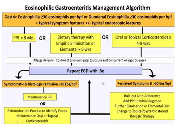
Treatment varies and is based on the severity of clinical features.
Avoid allergens or triggers
Common problem: Recurrence of symptoms associated with reintroduction of allergens such as certain foods
- Liquid diet (J Pediatr Gastroenterol Nutr 1996;23:81)
- Anti‐inflammatory drugs
- Systemic and topical steroids are considered the mainstay of treatment (Curr Allergy Asthma Rep 2005;5:259)
Some severe cases continue or fail to respond to dietary restrictions
Serous EGIDS usually responds better to steroids (Dig Dis Sci 2003;48:1013).
- Other steroid‐based therapies: Mast cell stabilizers (cromolyn sodium), mast cell secretion inhibitors (ketotifen), leukotriene receptor antagonists (montelukast), selective inhibitors of Th2, IL4 and IL5 cytokines (subplatast tosilate)
- Other targeted therapy options: Anti‐IgE monoclonal antibodies (omalizumab), anti‐IL5 antibodies (reslizumab and mepolizumab), and anti‐tissue activating factor alpha agents (infliximab)
- Fecal Microbiota Transplantation or FMT therapy (Fecal Microbiota Transplantation World J Gastroenterol 2014;20:16368)
Treatment includes identification of possible allergens and their elimination from the patient’s diet, leukotriene antagonists montelukast , or mast cell stabilizers ketotifen , systemic corticosteroids , ! important; azathioprine , and a liquid diet. It is difficult to deliver topical steroids to the gastric mucosa. The efficacy of cromoglycate preparations has not been established. In a study by Travis Piester et al. (2021) on gastritis and ulcer disease, eosinophilic gastroenteritis was often associated with allergies and atopic diseases. Eosinophilic gastritis often presents as an independent pathology in eosinophilic gastroenteritis rather than as a separate disease. Eosinophilic gastritis begins in the third decade of life, although 20% of cases may present before the age of 20;
Eosinophilic gastritis is also associated with other conditions such as irritable bowel syndrome, collagen vascular disease, bacterial and parasitic infections (including H. pylori ), drug injury, and drug sensitivity. Although eosinophilic gastritis can be seen in a variety of conditions, eosinophilia may be prominent in allergic and atopic conditions. In patients with eosinophilic gastritis, the medical history may include seasonal allergies, food sensitivities, eczema, and atopic dermatitis. The typical gastrointestinal clinical features are secondary to a combination of IgE and non‐IgE allergic reactions resulting from increased levels of interleukin 3 (IL‐3), IL‐5, and granulocyte‐monocyte colony‐stimulating factor, all of which are proinflammatory cytokines;
Eosinophilic gastritis may be mucosal (the most common subtype), muscular, serous, or a combination of these. The symptoms of eosinophilic gastritis are similar to those of other forms of gastritis. Cow’s milk, soy milk, eggs, and wheat are the most common allergens in children with allergic eosinophilic gastrointestinal disease, and the history suggests a relationship between food ingestion and symptoms of vomiting, hematemesis, irritability, and difficulty gaining weight. Young children may have anorexia and often refuse to eat. In rare cases, eosinophilic gastroenteritis may present with gastric obstruction due to swelling of mucosal folds when the stomach is empty. Anemia due to occult blood loss and hypoalbuminemia are common. If muscular or serous involvement occurs, abdominal pain and eosinophilic ascites may be present;
Endoscopic findings are also variable and nonspecific. Red hyperemia, pustularity, lacunae, ulceration, swollen gastric mucosal folds, and numerous scattered nodular lesions in the mucosa, especially in the antrum. These nonspecific findings emphasize the need for mucosal biopsy, which shows eosinophilic infiltration of the gastric mucosa and infiltration of lymphocytes, mast cells, and neutrophils. The histologic criteria for diagnosis are > 50 eosinophils/high‐power field in at least 5 fields, in the absence of other disorders with known eosinophilia. In the body, lesions are diffuse, while in the antrum smaller focal lesions predominate. If deeper layers are involved, endoscopic biopsy may not be diagnostic and endoscopic ultrasound or MRI may demonstrate thickening of the gastric wall;
Although a definitive diagnosis of food allergy requires allergen testing, a suspected diagnosis is based on response to allergen elimination and improvement in histologic appearance on endoscopy. When clinically present in young children, antigen presentation may occur within the first 24 months of life, although allergies to peanuts, tree nuts, and seafood may be present. Eosinophilic gastroenteritis is diagnosed based on the criteria proposed by Klein et al., which include gastrointestinal symptoms, tissue biopsy showing eosinophilic infiltration in one or more areas of the gastrointestinal tract, from the esophagus to the colon, or imaging evidence of peripheral eosinophilia, and absence of evidence of parasitic or extraintestinal disease. Only 50% of patients with eosinophilic gastroenteritis will have peripheral eosinophilia, which may be related to coexisting atopic disease without clinical gastrointestinal manifestations;
Targeted therapy includes removal of suspected or confirmed allergens, leukotriene receptor antagonists ( montelukast), mast cell stabilizers (ketotifen), systemic corticosteroids , azathioprine , or dietary modification. Gastric‐coating steroids are also a challenge. Symptomatic treatment may include acid suppression. Some reports of partial response are due to prolonged survival of eosinophils, and in part to increased IL‐5 levels.
In some severe cases, azathioprine and mycophenolate mofetil may be used and have been shown to be effective, especially in relapsed, steroid‐dependent cases. In young children, treatment includes a modified diet and water‐soluble protein to resolve symptoms and normalize biochemical markers (anemia and serum albumin) and endoscopic findings.
(Final)
Author: Dr. Huynh Hong Quang
(Quy Nhon Institute of Malaria, Parasitology and Entomology


