

Eosinophilic gastroenteritis is a group of gastrointestinal disorders with eosinophilia (EGIDs) including eosinophilic esophagitis, eosinophilic gastritis, eosinophilic gastroenteritis, eosinophilic enteritis, and eosinophilic colitis (EOS) according to criteria published in J Allergy Clin Immunol 2004.
Viêm dạ dày ruột non tăng bạch cầu ái toan là một rối loạn bệnh lý dạ dày ruột có tăng bạch cầu ái toan (Eosinophilic gastrointestinal disorders_EGIDs) gồm có viêm thực quản tăng BCAT, viêm dạ dày tăng BCAT, viêm ruột non tăng BCAT và viêm đại tràng tăng BCAT (eosinophilic esophagitis, eosinophilic gastritis, eosinophilic gastroenteritis, eosinophilic enteritis, eosinophilic colitis) theo tiêu chí đăng tải trên J Allergy Clin Immunol 2004.
However, eosinophilic esophagitis differs from eosinophilic gastroenterocolitis (EGEC) in all aspects and should be considered in its entirety when making the diagnosis. Eosinophilic gastroenterocolitis (EGEC) is an inflammatory disorder characterized by a high and predominant eosinophilic infiltration of the entire gastrointestinal tract without any associated cause or tissue with eosinophilic infiltration.
SOME IMPORTANT POINTS
The inflammatory process is characterized by abnormal infiltration of eosinophils into different segments of the gastrointestinal tract, in the absence of a known cause of eosinophilia (drug reactions, parasitic infections, and malignancies)
Can occur throughout the gastrointestinal tract, most commonly in the stomach, followed by the intestines and colon, with partial or multiple involvement of the gastrointestinal tract. Rarely in the biliary tract (Gut 1990;31:54)
Diagnosis requires the presence of: Gastrointestinal symptoms, tissue biopsy showing increased eosinophilia in the gastrointestinal tract. Exclusion of other causes of increased eosinophilia in tissues.
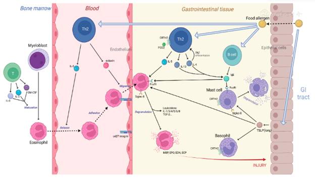
The cause is not fully understood. Some studies have suggested immune dysregulation or allergy because many patients have disorders because many patients with eosinophilic gastroenteritis disorders (EGIDs) are also associated with atopic dermatitis (Pediatr Neonatol 2011;52:272)
EGIDs can be classified into primary and secondary (Clin Rev Allergy Immunol 2016;50:175):
- Primary EGIDs are defined as an inflammatory process characterized by eosinophilic infiltration of various segments of the gastrointestinal tract, in the absence of previous causes of eosinophilia such as drug reactions, parasitic infections, or malignancies (J Allergy Clin Immunol 2004;113:11)
- EGIDs secondary to drug reactions, parasitic infections, bacterial infections, eosinophilic hyperinfective syndrome, Churg‐Strauss syndrome, celiac disease, inflammatory bowel disease, polyarteritis nodosa, and other connective tissue disorders.
CLINICAL CONTROVERSY
Nonspecific gastrointestinal symptoms: Abdominal pain, nausea, vomiting and diarrhea, malnutrition, growth retardation and weight loss;
Several symptoms may be common, depending on the depth of the gastrointestinal tract affected (J Allergy Clin Immunol 2004;113:11):
- Mucosal form: Abdominal pain, nausea, vomiting, diarrhea, bleeding, malabsorption, weight loss;
- Muscular: Intestinal obstruction and gastric obstruction;
- Serous type: Ascites fluid test shows increased BCAT.
Often present in patients with a history of atopic asthma, eczema, allergic rhinitis and increased serum IgE;
Some patients present with gross or microscopic blood in the stool (hematochezia), which can lead to significant anemia (Pediatr Neonatol 2011;52:272)
Rare cases of EGIDs have been associated with pancreatitis and duodenal ulceration or perforation (World J Gastroenterol 2009;15:2156, Endoscopy 2011;43:E358).
UPDATE ON HISTOPATHOLOGICAL IMAGES OF CYTOCYTOSIS OF THE DIGESTIVE SYSTEM
General
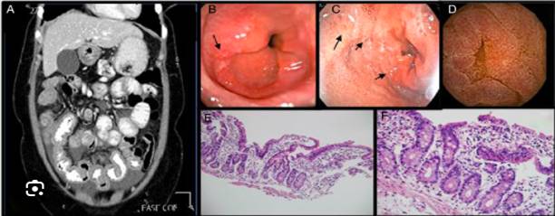
Nonspecific images on the mucosal surface, including thickened mucosa, red congestion, erosive ulcers and digestive ulcers, formation of white patches, edema, dilated blood vessels or formation of submucosal nodules.
Microscopic
Diagnosis requires confirmation by histopathological analysis of the stomach, small intestine, large intestine or biopsy ( Adv Anat Pathol 2011;18:335)
- BCAT distribution may be patchy (obtain multiple biopsies to confirm diagnosis)
Eosinophils can be seen throughout the gastrointestinal tract, except in the esophagus under normal physiological conditions they are an important part of the innate immune system of the digestive system;
- The normal range of BCAT numbers at different sites in the gastrointestinal tract also varies significantly between different patients (J Allergy Clin Immunol 2004;113:11);
- The highest concentrations of BCAT were found in the cecum and appendix (up to 30/high‐power field of view ( Mod Pathol 1996;9:110 , J Gastroenterol 2008;43:741 )
- There is a lack of consensus on the histological appearance of EGEC due to the rarity of the disease and the variability in the number of eosinophils, but it is characterized by a prominent eosinophilic infiltration of the gastrointestinal wall (mainly affected areas are the stomach and small intestine (Case Rep Gastroenterol 2013;7:293)
There is currently no cut‐off for the number of BCATs/high‐magnification microscopy to diagnose EGECs
Pathologists should discuss results if BCAT is more than expected
A recently proposed BCAT threshold is proposed here but is not a formal diagnostic criterion (Gastroenterol Clin North Am 2014;43:257):
- Stomach: ≥ 30 BCAT/microscopic field on 5 high magnification fields
- Duodenum: > 52 BCAT/high magnification field;
- Ileum: > 56 BCAT/high magnification field;
- Right colon: > 100 BCAT/high magnification field;
- Transverse colon and descending colon: 84 BCAT/ high magnification field;
- Sigmoid colon: > 64 BCAT/high magnification field .
BCAT distribution is also important evidence for EGEC regardless of BCAT number
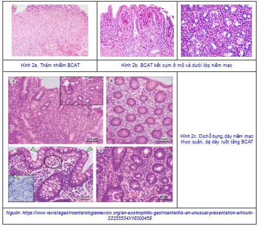
- The presence of eosinophils in cavities, crypts (intraepithelial eosinophils and eosinophilic gastrointestinal abscesses), eosinophilic clusters in the mucosa and eosinophils in the muscle are abnormal
In specimens excised from the pathology, total eosinophilic infiltration can be seen
May be accompanied by other inflammatory cells, especially mast cells
Other nonspecific changes include edema, loss of mucin, defects, reactivated epithelial changes (hyperplasia of surface mucous cells and ciliary hypertrophy), and mild structural disorganization
The area of increased eosinophilic inflammation may show extracellular eosinophilic deposition (Gastroenterology 1992;103:137)
Klein et al classified the disease based on the location of eosinophilic infiltration in different layers of the intestinal wall (depth of eosinophilic infiltration) into the mucosa, muscle, and serosa of the disease (Medicine (Baltimore) 1970;49:299):
Mucosal:
Most of the reports recorded in this class with 57‐100% of cases ( Gut 1990;31:54, Clin Gastroenterol Hepatol 2010;8:669)
Mucosal infiltration without involving the muscularis or serosa.
Muscle:
This is the second most commonly affected class
Predominant eosinophilic infiltration in the muscle layer
Causes thickening of the muscular layer, leading to obstruction of the stomach, small intestine or intussusception without increased BCAT ascites (World J Gastroenterol 2007;13:1758, Am Surg 1997;63:741)
Subserosal:
This form is uncommon estimated to account for only 9% in Japan and 13% in the United States ( Intern Med 1996;35:779)
Eosinophilic infiltration in the serosa
The main point of this category is increased BCAT abdominal effusion
Often accompanied by peripheral blood eosinophilia and peritonitis (Medicine (Baltimore) 1970;49:299)
There may be isolated localized ascites or ascites with characteristic symptoms of mucosal and muscular EGIDSs
Chang et al. saw simultaneous eosinophilic gastroenteritis in the muscular and serosal forms, indicating disease progression from the mucosal to the serosal form (Clin Gastroenterol Hepatol 2010;8:669).
Staining of clinical specimens (Major basic protein‐MBP;Allergy 2000;55:985)
Small intestine and stomach biopsy:
- Significant eosinophilic infiltration was identified in both the small intestinal and gastric mucosa
- Biopsy revealed diffuse eosinophilic infiltration (> 50/high‐power field) involving the mucosa and submucosa with intraepithelial eosinophils. No bacterial or parasitic findings, neutrophilic leukocytes, vasculitis, or granulomas were identified. On the basis of appropriate clinical findings, the findings suggest eosinophilic gastroenteritis.
- Clinical and pathologic correlation and further investigation may rule out secondary causes of eosinophilia.
(continute)
Author: Dr. Huynh Hong Quang
(Quy Nhon Institute of Malaria, Parasitology and Entomology)


